 Movie
Movie Controller


+ Open data

- Basic information
| Entry | Database: PDB / ID: 5bs8 | ||||||
|---|---|---|---|---|---|---|---|
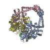
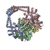
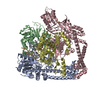
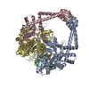
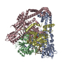
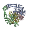
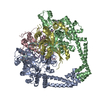
| Title | Crystal structure of a topoisomerase II complex | ||||||
 Components Components |
| ||||||
 Keywords Keywords | Isomerase/DNA / protein-DNA complex / topoisomerase II / Isomerase-DNA complex | ||||||
| Function / homology |  Function and homology information Function and homology informationDNA topoisomerase type II (double strand cut, ATP-hydrolyzing) complex / DNA negative supercoiling activity / DNA topoisomerase type II (double strand cut, ATP-hydrolyzing) activity / DNA topoisomerase (ATP-hydrolysing) / DNA topological change / peptidoglycan-based cell wall / DNA-templated DNA replication / chromosome / response to antibiotic / magnesium ion binding ...DNA topoisomerase type II (double strand cut, ATP-hydrolyzing) complex / DNA negative supercoiling activity / DNA topoisomerase type II (double strand cut, ATP-hydrolyzing) activity / DNA topoisomerase (ATP-hydrolysing) / DNA topological change / peptidoglycan-based cell wall / DNA-templated DNA replication / chromosome / response to antibiotic / magnesium ion binding / ATP hydrolysis activity / DNA binding / ATP binding / plasma membrane / cytoplasm Similarity search - Function | ||||||
| Biological species |   Mycobacterium tuberculosis (bacteria) Mycobacterium tuberculosis (bacteria)synthetic construct (others) | ||||||
| Method |  X-RAY DIFFRACTION / SYNCHROTRON / MOLECULAR REPLACEMENT / Resolution: 2.399 Å X-RAY DIFFRACTION / SYNCHROTRON / MOLECULAR REPLACEMENT / Resolution: 2.399 Å | ||||||
 Authors Authors | Blower, T.R. / Williamson, B.H. / Kerns, R.J. / Berger, J.M. | ||||||
 Citation Citation | Journal: Proc.Natl.Acad.Sci.USA / Year: 2016 Title: Crystal structure and stability of gyrase-fluoroquinolone cleaved complexes from Mycobacterium tuberculosis. Authors: Blower, T.R. / Williamson, B.H. / Kerns, R.J. / Berger, J.M. | ||||||
| History |
|
- Structure visualization
| Structure viewer | Molecule: MolmilJmol/JSmol |
|---|
- Downloads & links
-Download
| PDBx/mmCIF format | 5bs8.cif.gz | 956.6 KB | Display | PDBx/mmCIF format |
|---|---|---|---|---|
| PDB format | pdb5bs8.ent.gz | 804 KB | Display | PDB format |
| PDBx/mmJSON format | 5bs8.json.gz | Tree view | PDBx/mmJSON format | |
| Others |  Other downloads Other downloads |
-Validation report
| Arichive directory | https://data.pdbj.org/pub/pdb/validation_reports/bs/5bs8ftp://data.pdbj.org/pub/pdb/validation_reports/bs/5bs8 | HTTPS FTP |
|---|
-Related structure data
| Related structure data |  5btaC  5btcC  5btdC  5btfC  5btgC  5btiC  5btlC  5btnC  2xkkS C: citing same article ( S: Starting model for refinement |
|---|---|
| Similar structure data |


-Links
 PDBj
PDBj


- Assembly
| Deposited unit | 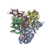
| ||||||||
|---|---|---|---|---|---|---|---|---|---|
| 1 |
| ||||||||
| Unit cell |
| ||||||||
| Details | The double-stranded DNAs are superposed copies of each other, overlaid in opposite orientations (because directionality could not be resolved). Therefore in reality, rather than in the model, the biological assembly would in fact be hexameric - four proteins and one double-stranded DNA. |
-Components
-DNA gyrase subunit ... , 2 types, 4 molecules ACBD
| #1: Protein | Mass: 56209.422 Da / Num. of mol.: 2 / Fragment: GyrA tower and C-gate domains Source method: isolated from a genetically manipulated source Source: (gene. exp.) Mycobacterium tuberculosis (strain ATCC 25618 / H37Rv) (bacteria)Strain: ATCC 25618 / H37Rv / Gene: gyrA, Rv0006, MTCY10H4.04 / Plasmid: pET-28b / Production host: #2: Protein | Mass: 28272.412 Da / Num. of mol.: 2 / Fragment: GyrB toprim domain Source method: isolated from a genetically manipulated source Source: (gene. exp.) Mycobacterium tuberculosis (strain ATCC 25618 / H37Rv) (bacteria)Strain: ATCC 25618 / H37Rv / Gene: gyrB, Rv0005, MTCY10H4.03 / Plasmid: pET-28b / Production host: |
|---|
-DNA substrate 24-mer ... , 2 types, 4 molecules EHFG
| #3: DNA chain | Mass: 7417.816 Da / Num. of mol.: 2 / Source method: obtained synthetically / Source: (synth.) synthetic construct (others) #4: DNA chain | Mass: 7319.739 Da / Num. of mol.: 2 / Source method: obtained synthetically / Source: (synth.) synthetic construct (others) |
|---|
-Non-polymers , 3 types, 409 molecules 


| #5: Chemical | ChemComp-MG /  Mass: 24.305 Da / Num. of mol.: 4 / Source method: obtained synthetically / Formula: Mg Mass: 24.305 Da / Num. of mol.: 4 / Source method: obtained synthetically / Formula: Mg#6: Chemical |  Mass: 401.431 Da / Num. of mol.: 2 / Source method: obtained synthetically / Formula: C21H24FN3O4 / Comment: antibiotic*YM Mass: 401.431 Da / Num. of mol.: 2 / Source method: obtained synthetically / Formula: C21H24FN3O4 / Comment: antibiotic*YM#7: Water | ChemComp-HOH / | Mass: 18.015 Da / Num. of mol.: 403 / Source method: isolated from a natural source / Formula: H2O |
|---|
-Details
| Has protein modification | Y |
|---|
-Experimental details
-Experiment
| Experiment | Method: X-RAY DIFFRACTION / Number of used crystals: 1 |
|---|
- Sample preparation
| Crystal | Density Matthews: 2.83 Å3/Da / Density % sol: 56.58 % / Description: Very thin planar crystals (<10 uM thick), up to 200 x 300 uM2 surface area |
|---|---|
| Crystal grow | Temperature: 294 K / Method: vapor diffusion, hanging drop / pH: 7 Details: 100 mM Tris-HCl, 13-15% PEG 4000, 200 mM MgCl2, 6.25% PEG 400 PH range: 7.0 to 8.5 |
-Data collection
| Diffraction | Mean temperature: 100 K |
|---|---|
| Diffraction source | Source: SYNCHROTRON / Site: APS  / Beamline: 24-ID-C / Wavelength: 0.979 Å / Beamline: 24-ID-C / Wavelength: 0.979 Å |
| Detector | Type: DECTRIS PILATUS 6M-F / Detector: PIXEL / Date: Jul 30, 2014 |
| Radiation | Monochromator: Si 220 filter / Protocol: SINGLE WAVELENGTH / Monochromatic (M) / Laue (L): M / Scattering type: x-ray |
| Radiation wavelength | Wavelength: 0.979 Å / Relative weight: 1 |
| Reflection | Resolution: 2.399→102.3 Å / Num. obs: 83492 / % possible obs: 97.68 % / Redundancy: 3.4 % / Rmerge(I) obs: 0.08799 / Net I/σ(I): 10.66 |
| Reflection shell | Resolution: 2.399→2.485 Å / Redundancy: 3.3 % / Mean I/σ(I) obs: 0.99 / Rejects: 0 / % possible all: 93.22 |
- Processing
| Software |
| |||||||||||||||||||||||||||||||||||||||||||||||||||||||||||||||||||||||||||||||||||||||||||||||||||||||||||||||||||||||||||||||||||||||||||||||||||||||||||||||||||||||||||||||||||||||||||||||||||||||||||||||||||||||||
|---|---|---|---|---|---|---|---|---|---|---|---|---|---|---|---|---|---|---|---|---|---|---|---|---|---|---|---|---|---|---|---|---|---|---|---|---|---|---|---|---|---|---|---|---|---|---|---|---|---|---|---|---|---|---|---|---|---|---|---|---|---|---|---|---|---|---|---|---|---|---|---|---|---|---|---|---|---|---|---|---|---|---|---|---|---|---|---|---|---|---|---|---|---|---|---|---|---|---|---|---|---|---|---|---|---|---|---|---|---|---|---|---|---|---|---|---|---|---|---|---|---|---|---|---|---|---|---|---|---|---|---|---|---|---|---|---|---|---|---|---|---|---|---|---|---|---|---|---|---|---|---|---|---|---|---|---|---|---|---|---|---|---|---|---|---|---|---|---|---|---|---|---|---|---|---|---|---|---|---|---|---|---|---|---|---|---|---|---|---|---|---|---|---|---|---|---|---|---|---|---|---|---|---|---|---|---|---|---|---|---|---|---|---|---|---|---|---|---|
| Refinement | Method to determine structure: MOLECULAR REPLACEMENT Starting model: 2XKK Resolution: 2.399→102.252 Å / SU ML: 0.44 / Cross valid method: FREE R-VALUE / σ(F): 1.89 / Phase error: 26.98 / Stereochemistry target values: ML
| |||||||||||||||||||||||||||||||||||||||||||||||||||||||||||||||||||||||||||||||||||||||||||||||||||||||||||||||||||||||||||||||||||||||||||||||||||||||||||||||||||||||||||||||||||||||||||||||||||||||||||||||||||||||||
| Solvent computation | Shrinkage radii: 0.9 Å / VDW probe radii: 1.11 Å / Solvent model: FLAT BULK SOLVENT MODEL | |||||||||||||||||||||||||||||||||||||||||||||||||||||||||||||||||||||||||||||||||||||||||||||||||||||||||||||||||||||||||||||||||||||||||||||||||||||||||||||||||||||||||||||||||||||||||||||||||||||||||||||||||||||||||
| Displacement parameters | Biso max: 319.72 Å2 / Biso mean: 77.0026 Å2 / Biso min: 27.31 Å2 | |||||||||||||||||||||||||||||||||||||||||||||||||||||||||||||||||||||||||||||||||||||||||||||||||||||||||||||||||||||||||||||||||||||||||||||||||||||||||||||||||||||||||||||||||||||||||||||||||||||||||||||||||||||||||
| Refinement step | Cycle: final / Resolution: 2.399→102.252 Å
| |||||||||||||||||||||||||||||||||||||||||||||||||||||||||||||||||||||||||||||||||||||||||||||||||||||||||||||||||||||||||||||||||||||||||||||||||||||||||||||||||||||||||||||||||||||||||||||||||||||||||||||||||||||||||
| Refine LS restraints |
| |||||||||||||||||||||||||||||||||||||||||||||||||||||||||||||||||||||||||||||||||||||||||||||||||||||||||||||||||||||||||||||||||||||||||||||||||||||||||||||||||||||||||||||||||||||||||||||||||||||||||||||||||||||||||
| LS refinement shell | Refine-ID: X-RAY DIFFRACTION / Total num. of bins used: 30
|
