 Movie
Movie Controller

[English] 日本語
 Yorodumi
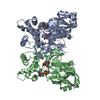
- PDB-5bre: Crystal structure of Trypanosoma cruzi glucokinase in complex wit... -
+ Open data

- Basic information
| Entry | Database: PDB / ID: 5bre | |||||||||
|---|---|---|---|---|---|---|---|---|---|---|
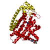
| Title | Crystal structure of Trypanosoma cruzi glucokinase in complex with inhibitor CBZ-GlcN | |||||||||
 Components Components | Glucokinase 1, putative | |||||||||
 Keywords Keywords | Transferase/Transferase Inhibitor / Transferase / Hexose Kinase / Transferase-Transferase Inhibitor complex | |||||||||
| Function / homology |  Function and homology information Function and homology informationglucokinase / glucokinase activity / D-glucose binding / glycolytic process / ATP binding Similarity search - Function | |||||||||
| Biological species |  | |||||||||
| Method |  X-RAY DIFFRACTION / SYNCHROTRON / MOLECULAR REPLACEMENT / molecular replacement / Resolution: 2.5 Å X-RAY DIFFRACTION / SYNCHROTRON / MOLECULAR REPLACEMENT / molecular replacement / Resolution: 2.5 Å | |||||||||
 Authors Authors | D'Antonio, E.L. / Perry, K. / Deinema, M.S. / Kearns, S.P. / Frey, T.A. | |||||||||
| Funding support |  United States, 2items United States, 2items
| |||||||||
 Citation Citation | Journal: Mol.Biochem.Parasitol. / Year: 2016 Title: Structure-based approach to the identification of a novel group of selective glucosamine analogue inhibitors of Trypanosoma cruzi glucokinase. Authors: D'Antonio, E.L. / Deinema, M.S. / Kearns, S.P. / Frey, T.A. / Tanghe, S. / Perry, K. / Roy, T.A. / Gracz, H.S. / Rodriguez, A. / D'Antonio, J. | |||||||||
| History |
|
- Structure visualization
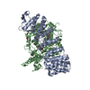
| Structure viewer | Molecule: MolmilJmol/JSmol |
|---|
- Downloads & links
-Download
| PDBx/mmCIF format | 5bre.cif.gz | 155.2 KB | Display | PDBx/mmCIF format |
|---|---|---|---|---|
| PDB format | pdb5bre.ent.gz | 120 KB | Display | PDB format |
| PDBx/mmJSON format | 5bre.json.gz | Tree view | PDBx/mmJSON format | |
| Others |  Other downloads Other downloads |
-Validation report
| Arichive directory | https://data.pdbj.org/pub/pdb/validation_reports/br/5breftp://data.pdbj.org/pub/pdb/validation_reports/br/5bre | HTTPS FTP |
|---|
-Related structure data
| Related structure data | 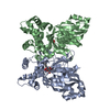 5brdC 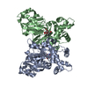 5brfC 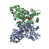 5brhC  2q2rS S: Starting model for refinement C: citing same article ( |
|---|---|
| Similar structure data |


-Links
 PDBj
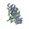
PDBj- Assembly
| Deposited unit | 
| ||||||||
|---|---|---|---|---|---|---|---|---|---|
| 1 |
| ||||||||
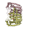
| Unit cell |
|
-Components
| #1: Protein | Mass: 42226.648 Da / Num. of mol.: 2 / Fragment: UNP residues 1-367 Source method: isolated from a genetically manipulated source Source: (gene. exp.) Strain: CL Brener / Gene: Tc00.1047053510187.100 / Plasmid: pET-28a(+) / Production host:  #2: Sugar |   Type: D-saccharide, beta linking / Mass: 313.303 Da / Num. of mol.: 2 / Source method: obtained synthetically / Formula: C14H19NO7 Type: D-saccharide, beta linking / Mass: 313.303 Da / Num. of mol.: 2 / Source method: obtained synthetically / Formula: C14H19NO7#3: Water | ChemComp-HOH / |  Mass: 18.015 Da / Num. of mol.: 4 / Source method: isolated from a natural source / Formula: H2O Mass: 18.015 Da / Num. of mol.: 4 / Source method: isolated from a natural source / Formula: H2O |
|---|
-Experimental details
-Experiment
| Experiment | Method: X-RAY DIFFRACTION / Number of used crystals: 1 |
|---|
- Sample preparation
| Crystal | Density Matthews: 2.54 Å3/Da / Density % sol: 51.64 % |
|---|---|
| Crystal grow | Temperature: 295 K / Method: vapor diffusion, sitting drop / pH: 7 Details: A glucose-free TcGlcK crystal was soaked in 1.0 mM CBZ-GlcN, 5.0% (v/v) DMSO, 0.1 M sodium citrate (pH 7.0), 15% (w/v) PEG 3,350 for 24 hours |
-Data collection
| Diffraction | Mean temperature: 100 K | |||||||||||||||||||||||||||||||||||||||||||||||||||||||||||||||||||||||||||||||||||||||||||||||||||
|---|---|---|---|---|---|---|---|---|---|---|---|---|---|---|---|---|---|---|---|---|---|---|---|---|---|---|---|---|---|---|---|---|---|---|---|---|---|---|---|---|---|---|---|---|---|---|---|---|---|---|---|---|---|---|---|---|---|---|---|---|---|---|---|---|---|---|---|---|---|---|---|---|---|---|---|---|---|---|---|---|---|---|---|---|---|---|---|---|---|---|---|---|---|---|---|---|---|---|---|---|
| Diffraction source | Source: SYNCHROTRON / Site: APS / Beamline: 24-ID-E / Wavelength: 0.97917 Å | |||||||||||||||||||||||||||||||||||||||||||||||||||||||||||||||||||||||||||||||||||||||||||||||||||
| Detector | Type: ADSC QUANTUM 315 / Detector: CCD / Date: Jul 26, 2014 / Details: LR-Design detector positioner | |||||||||||||||||||||||||||||||||||||||||||||||||||||||||||||||||||||||||||||||||||||||||||||||||||
| Radiation | Monochromator: Cryogenically-cooled single crystal Si(220) side bounce monochromator Protocol: SINGLE WAVELENGTH / Monochromatic (M) / Laue (L): M / Scattering type: x-ray | |||||||||||||||||||||||||||||||||||||||||||||||||||||||||||||||||||||||||||||||||||||||||||||||||||
| Radiation wavelength | Wavelength: 0.97917 Å / Relative weight: 1 | |||||||||||||||||||||||||||||||||||||||||||||||||||||||||||||||||||||||||||||||||||||||||||||||||||
| Reflection | Resolution: 2.5→50 Å / Num. obs: 28549 / % possible obs: 99.8 % / Redundancy: 3.7 % / Biso Wilson estimate: 52.98 Å2 / Rmerge(I) obs: 0.101 / Rpim(I) all: 0.062 / Rrim(I) all: 0.119 / Χ2: 1.122 / Net I/av σ(I): 13.262 / Net I/σ(I): 9.8 / Num. measured all: 105473 | |||||||||||||||||||||||||||||||||||||||||||||||||||||||||||||||||||||||||||||||||||||||||||||||||||
| Reflection shell | Diffraction-ID: 1 / Rejects: _
|
-Phasing
| Phasing | Method: molecular replacement |
|---|
- Processing
| Software |
| |||||||||||||||||||||||||||||||||||||||||||||||||||||||||||||||||||||||||||||
|---|---|---|---|---|---|---|---|---|---|---|---|---|---|---|---|---|---|---|---|---|---|---|---|---|---|---|---|---|---|---|---|---|---|---|---|---|---|---|---|---|---|---|---|---|---|---|---|---|---|---|---|---|---|---|---|---|---|---|---|---|---|---|---|---|---|---|---|---|---|---|---|---|---|---|---|---|---|---|
| Refinement | Method to determine structure: MOLECULAR REPLACEMENT Starting model: PDB entry 2Q2R Resolution: 2.5→27.427 Å / FOM work R set: 0.7902 / SU ML: 0.33 / Cross valid method: THROUGHOUT / σ(F): 1.36 / Phase error: 27.86 / Stereochemistry target values: ML
| |||||||||||||||||||||||||||||||||||||||||||||||||||||||||||||||||||||||||||||
| Solvent computation | Shrinkage radii: 0.9 Å / VDW probe radii: 1.11 Å / Solvent model: FLAT BULK SOLVENT MODEL | |||||||||||||||||||||||||||||||||||||||||||||||||||||||||||||||||||||||||||||
| Displacement parameters | Biso max: 134.44 Å2 / Biso mean: 78.49 Å2 / Biso min: 42.8 Å2 | |||||||||||||||||||||||||||||||||||||||||||||||||||||||||||||||||||||||||||||
| Refinement step | Cycle: final / Resolution: 2.5→27.427 Å
| |||||||||||||||||||||||||||||||||||||||||||||||||||||||||||||||||||||||||||||
| Refine LS restraints |
| |||||||||||||||||||||||||||||||||||||||||||||||||||||||||||||||||||||||||||||
| LS refinement shell | Refine-ID: X-RAY DIFFRACTION / Total num. of bins used: 10
|
