 Movie
Movie Controller

[English] 日本語
 Yorodumi
- PDB-5bnw: The active site of O-GlcNAc transferase imposes constraints on su... -
+ Open data

- Basic information
| Entry | Database: PDB / ID: 5bnw | ||||||
|---|---|---|---|---|---|---|---|
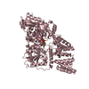
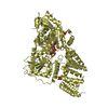
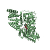
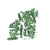
| Title | The active site of O-GlcNAc transferase imposes constraints on substrate sequence | ||||||
 Components Components |
| ||||||
 Keywords Keywords | TRANSFERASE / o-glcnac transferase / glycosyl transferase | ||||||
| Function / homology |  Function and homology information Function and homology informationnegative regulation of non-canonical inflammasome complex assembly / protein N-acetylglucosaminyltransferase complex / regulation of insulin receptor signaling pathway / protein O-GlcNAc transferase / protein localization to nuclear envelope / protein O-acetylglucosaminyltransferase activity / positive regulation of transcription from RNA polymerase II promoter by glucose / acetylglucosaminyltransferase activity / nuclear pore localization / regulation of Rac protein signal transduction ...negative regulation of non-canonical inflammasome complex assembly / protein N-acetylglucosaminyltransferase complex / regulation of insulin receptor signaling pathway / protein O-GlcNAc transferase / protein localization to nuclear envelope / protein O-acetylglucosaminyltransferase activity / positive regulation of transcription from RNA polymerase II promoter by glucose / acetylglucosaminyltransferase activity / nuclear pore localization / regulation of Rac protein signal transduction / regulation of necroptotic process / nuclear envelope organization / nuclear lamina / negative regulation of stem cell population maintenance / protein O-linked glycosylation / NSL complex / regulation of glycolytic process / regulation of gluconeogenesis / RIPK1-mediated regulated necrosis / intermediate filament / nuclear migration / Formation of WDR5-containing histone-modifying complexes / regulation of synapse assembly / Sin3-type complex / positive regulation of stem cell population maintenance / regulation of neurotransmitter receptor localization to postsynaptic specialization membrane / positive regulation of proteolysis / phosphatidylinositol-3,4,5-trisphosphate binding / histone acetyltransferase complex / hemopoiesis / positive regulation of lipid biosynthetic process / mitophagy / cell projection / positive regulation of TORC1 signaling / response to nutrient / negative regulation of proteasomal ubiquitin-dependent protein catabolic process / negative regulation of protein ubiquitination / negative regulation of cell migration / positive regulation of translation / circadian regulation of gene expression / cellular response to glucose stimulus / negative regulation of transforming growth factor beta receptor signaling pathway / protein processing / chromatin DNA binding / response to insulin / structural constituent of cytoskeleton / Regulation of necroptotic cell death / mitochondrial membrane / UCH proteinases / positive regulation of cold-induced thermogenesis / HATs acetylate histones / heterochromatin formation / chromatin organization / nuclear membrane / apoptotic process / regulation of transcription by RNA polymerase II / positive regulation of DNA-templated transcription / glutamatergic synapse / negative regulation of transcription by RNA polymerase II / signal transduction / positive regulation of transcription by RNA polymerase II / protein-containing complex / nucleoplasm / identical protein binding / nucleus / plasma membrane / cytosol Similarity search - Function | ||||||
| Biological species |  Homo sapiens (human) Homo sapiens (human) | ||||||
| Method |  X-RAY DIFFRACTION / SYNCHROTRON / MOLECULAR REPLACEMENT / Resolution: 2.4 Å X-RAY DIFFRACTION / SYNCHROTRON / MOLECULAR REPLACEMENT / Resolution: 2.4 Å | ||||||
 Authors Authors | Pathak, S. / Alonso, J. / Schimpl, M. / Rafie, K. / Blair, D.E. / Borodkin, V.S. / Albarbarawi, O. / van Aalten, D.M.F. | ||||||
| Funding support |  United Kingdom, 1items United Kingdom, 1items
| ||||||
 Citation Citation | Journal: Nat.Struct.Mol.Biol. / Year: 2015 Title: The active site of O-GlcNAc transferase imposes constraints on substrate sequence. Authors: Pathak, S. / Alonso, J. / Schimpl, M. / Rafie, K. / Blair, D.E. / Borodkin, V.S. / Schuttelkopf, A.W. / Albarbarawi, O. / van Aalten, D.M. | ||||||
| History |
|
- Structure visualization
| Structure viewer | Molecule: MolmilJmol/JSmol |
|---|
- Downloads & links
-Download
| PDBx/mmCIF format | 5bnw.cif.gz | 156.8 KB | Display | PDBx/mmCIF format |
|---|---|---|---|---|
| PDB format | pdb5bnw.ent.gz | 119.2 KB | Display | PDB format |
| PDBx/mmJSON format | 5bnw.json.gz | Tree view | PDBx/mmJSON format | |
| Others |  Other downloads Other downloads |
-Validation report
| Arichive directory | https://data.pdbj.org/pub/pdb/validation_reports/bn/5bnwftp://data.pdbj.org/pub/pdb/validation_reports/bn/5bnw | HTTPS FTP |
|---|
-Related structure data
| Related structure data | 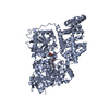 4xi9C 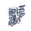 4xifC 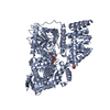 5c1dC  3pe4S S: Starting model for refinement C: citing same article ( |
|---|---|
| Similar structure data |


-Links
 PDBj
PDBj


- Assembly
| Deposited unit | 
| ||||||||
|---|---|---|---|---|---|---|---|---|---|
| 1 |
| ||||||||
| Unit cell |
|
-Components
| #1: Protein | Mass: 80974.508 Da / Num. of mol.: 1 / Fragment: UNP residues 197-915 Source method: isolated from a genetically manipulated source Source: (gene. exp.) Homo sapiens (human) / Gene: OGT / Production host:  |
|---|---|
| #2: Protein/peptide | Mass: 1346.531 Da / Num. of mol.: 1 / Source method: obtained synthetically / Source: (synth.) Homo sapiens (human) / References: UniProt: Q03252*PLUS |
| #3: Chemical | ChemComp-12V / (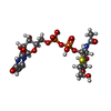  Mass: 623.419 Da / Num. of mol.: 1 / Source method: obtained synthetically / Formula: C17H27N3O16P2S Mass: 623.419 Da / Num. of mol.: 1 / Source method: obtained synthetically / Formula: C17H27N3O16P2S |
| #4: Water | ChemComp-HOH /  Mass: 18.015 Da / Num. of mol.: 78 / Source method: isolated from a natural source / Formula: H2O Mass: 18.015 Da / Num. of mol.: 78 / Source method: isolated from a natural source / Formula: H2O |
-Experimental details
-Experiment
| Experiment | Method: X-RAY DIFFRACTION |
|---|
- Sample preparation
| Crystal | Density Matthews: 2.21 Å3/Da / Density % sol: 44.34 % |
|---|---|
| Crystal grow | Temperature: 294 K / Method: vapor diffusion, hanging drop / pH: 6.4 Details: 1.3 M DL-Malic acid pH 6.4, 0.1 M Bis-Tris propane pH 6.4 supplemented with crystal seeds grown out of the same condition. |
-Data collection
| Diffraction | Mean temperature: 100 K |
|---|---|
| Diffraction source | Source: SYNCHROTRON / Site: Diamond / Beamline: I04-1 / Wavelength: 0.92 Å |
| Detector | Type: PSI PILATUS 6M / Detector: PIXEL / Date: May 9, 2015 / Details: Pilatus 2M Detector |
| Radiation | Protocol: SINGLE WAVELENGTH / Monochromatic (M) / Laue (L): M / Scattering type: x-ray |
| Radiation wavelength | Wavelength: 0.92 Å / Relative weight: 1 |
| Reflection | Resolution: 2.4→30 Å / Num. all: 263631 / Num. obs: 40463 / % possible obs: 99.9 % / Redundancy: 6.5 % / Rmerge(I) obs: 0.104 / Net I/σ(I): 11.8 |
| Reflection shell | Resolution: 2.4→2.53 Å / Redundancy: 6.7 % / Rmerge(I) obs: 0.719 / Mean I/σ(I) obs: 2.4 / % possible all: 100 |
- Processing
| Software |
| ||||||||||||||||||||||||||||||||||||||||||||||||||||||||||||||||||||||||||||||||||||||||||||||||||||||||||||||||||||||||||||||||||||||||||||||||||||||||||||||||||||||||||||||||||||||
|---|---|---|---|---|---|---|---|---|---|---|---|---|---|---|---|---|---|---|---|---|---|---|---|---|---|---|---|---|---|---|---|---|---|---|---|---|---|---|---|---|---|---|---|---|---|---|---|---|---|---|---|---|---|---|---|---|---|---|---|---|---|---|---|---|---|---|---|---|---|---|---|---|---|---|---|---|---|---|---|---|---|---|---|---|---|---|---|---|---|---|---|---|---|---|---|---|---|---|---|---|---|---|---|---|---|---|---|---|---|---|---|---|---|---|---|---|---|---|---|---|---|---|---|---|---|---|---|---|---|---|---|---|---|---|---|---|---|---|---|---|---|---|---|---|---|---|---|---|---|---|---|---|---|---|---|---|---|---|---|---|---|---|---|---|---|---|---|---|---|---|---|---|---|---|---|---|---|---|---|---|---|---|---|
| Refinement | Method to determine structure: MOLECULAR REPLACEMENT Starting model: 3PE4 Resolution: 2.4→30 Å / Cor.coef. Fo:Fc: 0.957 / Cor.coef. Fo:Fc free: 0.926 / SU B: 7.831 / SU ML: 0.174 / Cross valid method: THROUGHOUT / ESU R: 0.274 / ESU R Free: 0.22 / Stereochemistry target values: MAXIMUM LIKELIHOOD / Details: HYDROGENS HAVE BEEN ADDED IN THE RIDING POSITIONS
| ||||||||||||||||||||||||||||||||||||||||||||||||||||||||||||||||||||||||||||||||||||||||||||||||||||||||||||||||||||||||||||||||||||||||||||||||||||||||||||||||||||||||||||||||||||||
| Solvent computation | Ion probe radii: 0.8 Å / Shrinkage radii: 0.8 Å / VDW probe radii: 1.2 Å / Solvent model: MASK | ||||||||||||||||||||||||||||||||||||||||||||||||||||||||||||||||||||||||||||||||||||||||||||||||||||||||||||||||||||||||||||||||||||||||||||||||||||||||||||||||||||||||||||||||||||||
| Displacement parameters | Biso mean: 45.84 Å2
| ||||||||||||||||||||||||||||||||||||||||||||||||||||||||||||||||||||||||||||||||||||||||||||||||||||||||||||||||||||||||||||||||||||||||||||||||||||||||||||||||||||||||||||||||||||||
| Refinement step | Cycle: 1 / Resolution: 2.4→30 Å
| ||||||||||||||||||||||||||||||||||||||||||||||||||||||||||||||||||||||||||||||||||||||||||||||||||||||||||||||||||||||||||||||||||||||||||||||||||||||||||||||||||||||||||||||||||||||
| Refine LS restraints |
|