 Movie
Movie Controller


+ Open data

- Basic information
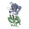
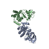
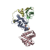
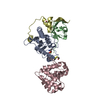
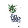
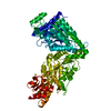
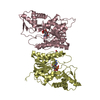
| Entry | Database: PDB / ID: 5b6b | ||||||
|---|---|---|---|---|---|---|---|
| Title | Complex of LATS1 and phosphomimetic MOB1b | ||||||
 Components Components |
| ||||||
 Keywords Keywords | METAL BINDING PROTEIN/SIGNALING PROTEIN / MOB1 LATS1 Hippo pathway / METAL BINDING PROTEIN-SIGNALING PROTEIN complex | ||||||
| Function / homology |  Function and homology information Function and homology informationinner cell mass cell fate commitment / inner cell mass cellular morphogenesis / regulation of protein autophosphorylation / Signaling by Hippo / negative regulation of cyclin-dependent protein serine/threonine kinase activity / regulation of ubiquitin-dependent protein catabolic process / regulation of transforming growth factor beta receptor signaling pathway / mammary gland epithelial cell differentiation / positive regulation of hippo signaling / regulation of intracellular estrogen receptor signaling pathway ...inner cell mass cell fate commitment / inner cell mass cellular morphogenesis / regulation of protein autophosphorylation / Signaling by Hippo / negative regulation of cyclin-dependent protein serine/threonine kinase activity / regulation of ubiquitin-dependent protein catabolic process / regulation of transforming growth factor beta receptor signaling pathway / mammary gland epithelial cell differentiation / positive regulation of hippo signaling / regulation of intracellular estrogen receptor signaling pathway / sister chromatid segregation / regulation of actin filament polymerization / regulation of postsynaptic density assembly / regulation of organ growth / hippo signaling / NLRP3 inflammasome complex assembly / kinase activator activity / negative regulation of protein localization to nucleus / positive regulation of NLRP3 inflammasome complex assembly / microtubule organizing center / keratinocyte differentiation / regulation of protein-containing complex assembly / protein kinase activator activity / hormone-mediated signaling pathway / nuclear estrogen receptor binding / protein serine/threonine kinase activator activity / G1/S transition of mitotic cell cycle / negative regulation of canonical Wnt signaling pathway / G2/M transition of mitotic cell cycle / kinase binding / spindle pole / intracellular protein localization / midbody / protein phosphorylation / non-specific serine/threonine protein kinase / postsynapse / positive regulation of apoptotic process / protein serine kinase activity / cell division / protein serine/threonine kinase activity / centrosome / nucleolus / protein kinase binding / glutamatergic synapse / magnesium ion binding / nucleoplasm / ATP binding / metal ion binding / nucleus / cytosol / cytoplasm Similarity search - Function | ||||||
| Biological species |  | ||||||
| Method |  X-RAY DIFFRACTION / SYNCHROTRON / MOLECULAR REPLACEMENT / Resolution: 3.536 Å X-RAY DIFFRACTION / SYNCHROTRON / MOLECULAR REPLACEMENT / Resolution: 3.536 Å | ||||||
 Authors Authors | KIM, S.-Y. / Tachioka, Y. / Mori, T. / Hakoshima, T. | ||||||
 Citation Citation | Journal: Sci Rep / Year: 2016 Title: Structural basis for autoinhibition and its relief of MOB1 in the Hippo pathway Authors: Kim, S.Y. / Tachioka, Y. / Mori, T. / Hakoshima, T. | ||||||
| History |
|
- Structure visualization
| Structure viewer | Molecule: MolmilJmol/JSmol |
|---|
- Downloads & links
-Download
| PDBx/mmCIF format | 5b6b.cif.gz | 416.8 KB | Display | PDBx/mmCIF format |
|---|---|---|---|---|
| PDB format | pdb5b6b.ent.gz | 341.5 KB | Display | PDB format |
| PDBx/mmJSON format | 5b6b.json.gz | Tree view | PDBx/mmJSON format | |
| Others |  Other downloads Other downloads |
-Validation report
| Arichive directory | https://data.pdbj.org/pub/pdb/validation_reports/b6/5b6bftp://data.pdbj.org/pub/pdb/validation_reports/b6/5b6b | HTTPS FTP |
|---|
-Related structure data
| Related structure data | 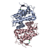 5b5vC 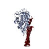 5b5wSC C: citing same article ( S: Starting model for refinement |
|---|---|
| Similar structure data |


-Links
 PDBj
PDBj


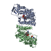
- Assembly
| Deposited unit | 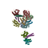
| ||||||||
|---|---|---|---|---|---|---|---|---|---|
| 1 | 
| ||||||||
| 2 | 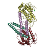
| ||||||||
| 3 | 
| ||||||||
| 4 | 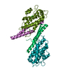
| ||||||||
| Unit cell |
|
-Components
| #1: Protein | Mass: 25304.953 Da / Num. of mol.: 8 / Mutation: T35D, T22D Source method: isolated from a genetically manipulated source Source: (gene. exp.)  #2: Protein | Mass: 10379.002 Da / Num. of mol.: 8 / Fragment: UNP residues 621-703 Source method: isolated from a genetically manipulated source Source: (gene. exp.) References: UniProt: Q8BYR2, non-specific serine/threonine protein kinase #3: Chemical | ChemComp-CL /   Mass: 35.453 Da / Num. of mol.: 7 / Source method: obtained synthetically / Formula: Cl Mass: 35.453 Da / Num. of mol.: 7 / Source method: obtained synthetically / Formula: Cl#4: Chemical | ChemComp-ZN /   Mass: 65.409 Da / Num. of mol.: 8 / Source method: obtained synthetically / Formula: Zn Mass: 65.409 Da / Num. of mol.: 8 / Source method: obtained synthetically / Formula: Zn |
|---|
-Experimental details
-Experiment
| Experiment | Method: X-RAY DIFFRACTION / Number of used crystals: 1 |
|---|
- Sample preparation
| Crystal | Density Matthews: 2.84 Å3/Da / Density % sol: 56.62 % |
|---|---|
| Crystal grow | Temperature: 277 K / Method: vapor diffusion, hanging drop / Details: PEG 3350, sodium sulfate |
-Data collection
| Diffraction | Mean temperature: 100 K |
|---|---|
| Diffraction source | Source: SYNCHROTRON / Site: SPring-8  / Beamline: BL44XU / Wavelength: 0.9 Å / Beamline: BL44XU / Wavelength: 0.9 Å |
| Detector | Type: RAYONIX MX300HE / Detector: CCD / Date: Nov 12, 2014 |
| Radiation | Protocol: SINGLE WAVELENGTH / Monochromatic (M) / Laue (L): M / Scattering type: x-ray |
| Radiation wavelength | Wavelength: 0.9 Å / Relative weight: 1 |
| Reflection | Resolution: 3.5→50 Å / Num. obs: 41452 / % possible obs: 100 % / Redundancy: 7.5 % / Rmerge(I) obs: 0.147 / Net I/σ(I): 19.9 |
| Reflection shell | Resolution: 3.56→30 Å |
- Processing
| Software |
| |||||||||||||||||||||||||||||||||||||||||||||||||||||||||||||||||||||||||||||||||||||||||||||||||||||||||
|---|---|---|---|---|---|---|---|---|---|---|---|---|---|---|---|---|---|---|---|---|---|---|---|---|---|---|---|---|---|---|---|---|---|---|---|---|---|---|---|---|---|---|---|---|---|---|---|---|---|---|---|---|---|---|---|---|---|---|---|---|---|---|---|---|---|---|---|---|---|---|---|---|---|---|---|---|---|---|---|---|---|---|---|---|---|---|---|---|---|---|---|---|---|---|---|---|---|---|---|---|---|---|---|---|---|---|
| Refinement | Method to determine structure: MOLECULAR REPLACEMENT Starting model: 5B5W Resolution: 3.536→48.644 Å / SU ML: 0.41 / Cross valid method: FREE R-VALUE / σ(F): 1.34 / Phase error: 28.25 / Stereochemistry target values: ML
| |||||||||||||||||||||||||||||||||||||||||||||||||||||||||||||||||||||||||||||||||||||||||||||||||||||||||
| Solvent computation | Shrinkage radii: 0.9 Å / VDW probe radii: 1.11 Å / Solvent model: FLAT BULK SOLVENT MODEL | |||||||||||||||||||||||||||||||||||||||||||||||||||||||||||||||||||||||||||||||||||||||||||||||||||||||||
| Refinement step | Cycle: LAST / Resolution: 3.536→48.644 Å
| |||||||||||||||||||||||||||||||||||||||||||||||||||||||||||||||||||||||||||||||||||||||||||||||||||||||||
| Refine LS restraints |
| |||||||||||||||||||||||||||||||||||||||||||||||||||||||||||||||||||||||||||||||||||||||||||||||||||||||||
| LS refinement shell |
|
