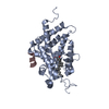
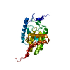
Entry Database : PDB / ID : 5b5bTitle Crystal structure of VDR-LBD complexed with 2-methylidene-26,27-diphenyl-19-nor-1,25-dihydroxyvitamin D3 Mediator of RNA polymerase II transcription subunit 1 Vitamin D3 receptor Keywords / / / / / / Function / homology Function Domain/homology Component
/ / / / / / / / / / / / / / / / / / / / / / / / / / / / / / / / / / / / / / / / / / / / / / / / / / / / / / / / / / / / / / / / / / / / / / / / / / / / / / / / / / / / / / / / / / / / / / / / / / / / / / / / / / / / / / / / / / / / / / / / / / / / / / / / / / / / / / / / / / Biological species Rattus norvegicus (Norway rat)Homo sapiens (human)Method / / Resolution : 2 Å Authors Kato, A. / Itoh, T. / Yamamoto, K. Journal : Bioconjug.Chem. / Year : 2016Title : Helix12-Stabilization Antagonist of Vitamin D ReceptorAuthors : Kato, A. / Itoh, T. / Anami, Y. / Egawa, D. / Yamamoto, K. History Deposition May 2, 2016 Deposition site / Processing site Revision 1.0 Jun 29, 2016 Provider / Type Revision 1.1 Aug 3, 2016 Group Revision 1.2 Feb 26, 2020 Group / Database references / Derived calculationsCategory / diffrn_source / pdbx_struct_oper_listItem / _diffrn_source.pdbx_synchrotron_site / _pdbx_struct_oper_list.symmetry_operationRevision 1.3 Mar 20, 2024 Group / Database references / Category / chem_comp_bond / database_2Item / _database_2.pdbx_database_accession
Show all Show less
 Movie
Movie Controller

 Yorodumi
 Open data
 Basic information
 Components
 Keywords
 Function and homology information
 Homo sapiens (human)
 X-RAY DIFFRACTION /
  Authors
 Citation
 Structure visualization
 Downloads & links
 Other downloads
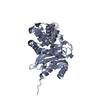


 PDBj
PDBj


 Assembly
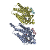


 Mass: 568.828 Da / Num. of mol.: 2 / Source method: obtained synthetically / Formula: C39H52O3
 Mass: 18.015 Da / Num. of mol.: 306 / Source method: isolated from a natural source / Formula: H2O
 Sample preparation
 / Beamline: AR-NW12A / Wavelength: 1 Å
 Processing