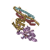
[English] 日本語
Yorodumi
- PDB-5axh: Crystal structure of thermophilic dextranase from Thermoanaerobac... -

+
Open data


ID or keywords:

Loading...

-
Basic information

Entry
Database: PDB / ID: 5axh
TitleCrystal structure of thermophilic dextranase from Thermoanaerobacter pseudethanolicus, D312G mutant in complex with isomaltohexaose
ComponentsDextranase
KeywordsHYDROLASE / glycoside hydrolase family 66
Function / homology
Function and homology information


dextranase / dextranase activity / metabolic process / membrane
Similarity search - Function
Glycosyl hydrolase family 66 / Glycosyl hydrolase family 66 / Golgi alpha-mannosidase II / Glycosyl hydrolase, all-beta / Glycosidases / TIM Barrel / Alpha-Beta Barrel / Immunoglobulin-like fold / Immunoglobulin-like / Sandwich ...Glycosyl hydrolase family 66 / Glycosyl hydrolase family 66 / Golgi alpha-mannosidase II / Glycosyl hydrolase, all-beta / Glycosidases / TIM Barrel / Alpha-Beta Barrel / Immunoglobulin-like fold / Immunoglobulin-like / Sandwich / Mainly Beta / Alpha Beta
Similarity search - Domain/homology
PHOSPHATE ION / Dextranase
Similarity search - Component
Biological speciesThermoanaerobacter pseudethanolicus ATCC 33223 (bacteria)
MethodX-RAY DIFFRACTION / SYNCHROTRON / MOLECULAR REPLACEMENT / Resolution: 2.2 Å
AuthorsSuzuki, N. / Kishine, N. / Fujimoto, Z. / Sakurai, M. / Momma, M. / Ko, J.A. / Nam, S.H. / Kimura, A. / Kim, Y.M.
Funding support Japan, Korea, Republic Of, 2items
OrganizationGrant numberCountry
Ministry of Agriculture, Forestry and Fisheries25001A Japan
National Research Foundation of Korea2010-0020141 Korea, Republic Of
CitationJournal: J.Biochem. / Year: 2016
Title: Crystal structure of thermophilic dextranase from Thermoanaerobacter pseudethanolicus
Authors: Suzuki, N. / Kishine, N. / Fujimoto, Z. / Sakurai, M. / Momma, M. / Ko, J.A. / Nam, S.H. / Kimura, A. / Kim, Y.M.
History
DepositionJul 29, 2015Deposition site: PDBJ / Processing site: PDBJ
Revision 1.0Nov 11, 2015Provider: repository / Type: Initial release
Revision 1.1Mar 9, 2016Group: Database references
Revision 1.2Feb 26, 2020Group: Data collection / Derived calculations / Category: chem_comp / diffrn_source / pdbx_struct_oper_list
Item: _chem_comp.type / _diffrn_source.pdbx_synchrotron_site / _pdbx_struct_oper_list.symmetry_operation
Revision 2.0Jul 29, 2020Group: Atomic model / Data collection ...Atomic model / Data collection / Derived calculations / Structure summary
Category: atom_site / chem_comp ...atom_site / chem_comp / entity / pdbx_branch_scheme / pdbx_chem_comp_identifier / pdbx_entity_branch / pdbx_entity_branch_descriptor / pdbx_entity_branch_link / pdbx_entity_branch_list / pdbx_entity_nonpoly / pdbx_nonpoly_scheme / pdbx_struct_assembly_gen / struct_asym / struct_conn / struct_site / struct_site_gen
Item: _atom_site.B_iso_or_equiv / _atom_site.Cartn_x ..._atom_site.B_iso_or_equiv / _atom_site.Cartn_x / _atom_site.Cartn_y / _atom_site.Cartn_z / _atom_site.auth_asym_id / _atom_site.auth_atom_id / _atom_site.auth_comp_id / _atom_site.auth_seq_id / _atom_site.label_asym_id / _atom_site.label_atom_id / _atom_site.label_comp_id / _atom_site.label_entity_id / _atom_site.type_symbol / _chem_comp.name / _entity.formula_weight / _entity.pdbx_description / _entity.pdbx_number_of_molecules / _entity.src_method / _entity.type / _pdbx_struct_assembly_gen.asym_id_list / _struct_conn.pdbx_dist_value / _struct_conn.ptnr1_auth_asym_id / _struct_conn.ptnr1_auth_comp_id / _struct_conn.ptnr1_auth_seq_id / _struct_conn.ptnr1_label_asym_id / _struct_conn.ptnr1_label_atom_id / _struct_conn.ptnr1_label_comp_id / _struct_conn.ptnr2_auth_asym_id / _struct_conn.ptnr2_auth_comp_id / _struct_conn.ptnr2_auth_seq_id / _struct_conn.ptnr2_label_asym_id / _struct_conn.ptnr2_label_atom_id / _struct_conn.ptnr2_label_comp_id
Description: Carbohydrate remediation / Provider: repository / Type: Remediation
Revision 2.1Nov 8, 2023Group: Data collection / Database references ...Data collection / Database references / Derived calculations / Refinement description / Structure summary
Category: chem_comp / chem_comp_atom ...chem_comp / chem_comp_atom / chem_comp_bond / database_2 / pdbx_initial_refinement_model / struct_conn
Item: _chem_comp.pdbx_synonyms / _database_2.pdbx_DOI ..._chem_comp.pdbx_synonyms / _database_2.pdbx_DOI / _database_2.pdbx_database_accession / _struct_conn.pdbx_leaving_atom_flag

-
Structure visualization

Structure viewerMolecule:
MolmilJmol/JSmol

Downloads & links

-
Assembly

Deposited unit
A: Dextranase
B: Dextranase
hetero molecules


Theoretical massNumber of molelcules
Total (without water)144,87610
Polymers142,6552
Non-polymers2,2228
Water10,701594
1


  • Idetical with deposited unit
  • defined by author&software
TypeNameSymmetry operationNumber
identity operation1_555x,y,z1
Buried area7160 Å2
ΔGint5 kcal/mol
Surface area43200 Å2
MethodPISA
Unit cell
Length a, b, c (Å)78.334, 99.179, 169.254
Angle α, β, γ (deg.)90.00, 90.00, 90.00
Int Tables number19
Space group name H-MP212121

-
Components

-
Protein , 1 types, 2 molecules AB

#1: Protein Dextranase /


Mass: 71327.484 Da / Num. of mol.: 2 / Fragment: UNP residues 3-610 / Mutation: D312G
Source method: isolated from a genetically manipulated source
Source: (gene. exp.) Thermoanaerobacter pseudethanolicus ATCC 33223 (bacteria)
Strain: ATCC 33223 / Gene: Teth39_0264 / Plasmid: pET23d / Production host: Escherichia coli BL21(DE3) (bacteria) / Strain (production host): BL21(DE3) / References: UniProt: B0KBZ7, dextranase

-
Sugars , 2 types, 2 molecules

#2: Polysaccharide alpha-D-glucopyranose-(1-6)-alpha-D-glucopyranose-(1-6)-alpha-D-glucopyranose-(1-6)-alpha-D-glucopyranose


Type: oligosaccharide / Mass: 666.578 Da / Num. of mol.: 1
Source method: isolated from a genetically manipulated source
DescriptorTypeProgram
DGlcpa1-6DGlcpa1-6DGlcpa1-6DGlcpa1-ROHGlycam Condensed SequenceGMML 1.0
WURCS=2.0/1,4,3/[a2122h-1a_1-5]/1-1-1-1/a6-b1_b6-c1_c6-d1WURCSPDB2Glycan 1.1.0
[][a-D-Glcp]{[(6+1)][a-D-Glcp]{[(6+1)][a-D-Glcp]{[(6+1)][a-D-Glcp]{}}}}LINUCSPDB-CARE
#3: Polysaccharide alpha-D-glucopyranose-(1-6)-alpha-D-glucopyranose-(1-6)-alpha-D-glucopyranose-(1-6)-alpha-D- ...alpha-D-glucopyranose-(1-6)-alpha-D-glucopyranose-(1-6)-alpha-D-glucopyranose-(1-6)-alpha-D-glucopyranose-(1-6)-alpha-D-glucopyranose-(1-6)-beta-D-glucopyranose


Type: oligosaccharide / Mass: 990.860 Da / Num. of mol.: 1
Source method: isolated from a genetically manipulated source
DescriptorTypeProgram
DGlcpa1-6DGlcpa1-6DGlcpa1-6DGlcpa1-6DGlcpa1-6DGlcpb1-ROHGlycam Condensed SequenceGMML 1.0
WURCS=2.0/2,6,5/[a2122h-1b_1-5][a2122h-1a_1-5]/1-2-2-2-2-2/a6-b1_b6-c1_c6-d1_d6-e1_e6-f1WURCSPDB2Glycan 1.1.0
[][b-D-Glcp]{[(6+1)][a-D-Glcp]{[(6+1)][a-D-Glcp]{[(6+1)][a-D-Glcp]{[(6+1)][a-D-Glcp]{[(6+1)][a-D-Glcp]{}}}}}}LINUCSPDB-CARE

-
Non-polymers , 3 types, 600 molecules

#4: Chemical
ChemComp-PO4 / PHOSPHATE ION / Phosphate


Mass: 94.971 Da / Num. of mol.: 4 / Source method: obtained synthetically / Formula: PO4
#5: Chemical ChemComp-GOL / GLYCEROL / GLYCERIN / PROPANE-1,2,3-TRIOL / Glycerol


Mass: 92.094 Da / Num. of mol.: 2 / Source method: obtained synthetically / Formula: C3H8O3
#6: Water ChemComp-HOH / water / Water


Mass: 18.015 Da / Num. of mol.: 594 / Source method: isolated from a natural source / Formula: H2O

-
Experimental details

-
Experiment

ExperimentMethod: X-RAY DIFFRACTION

-
Sample preparation

CrystalDensity Matthews: 2.42 Å3/Da / Density % sol: 49 % / Description: Rod
Crystal growTemperature: 293 K / Method: vapor diffusion, sitting drop / pH: 7.8
Details: 0.12 M sodium dihydrogenphosphate dihydrate, 1.68 M potassium phosphate dibasic

-
Data collection

DiffractionMean temperature: 95 K
Diffraction sourceSource: SYNCHROTRON / Site: Photon Factory / Beamline: BL-5A / Wavelength: 1 Å
DetectorType: ADSC QUANTUM 315 / Detector: CCD / Date: Jun 3, 2014
RadiationMonochromator: Si(111) / Protocol: SINGLE WAVELENGTH / Monochromatic (M) / Laue (L): M / Scattering type: x-ray
Radiation wavelengthWavelength: 1 Å / Relative weight: 1
ReflectionResolution: 2.2→38.92 Å / Num. obs: 67398 / % possible obs: 99.5 % / Observed criterion σ(F): 0 / Observed criterion σ(I): -3 / Redundancy: 13.7 % / Rmerge(I) obs: 0.127 / Rsym value: 0.122 / Net I/σ(I): 22.2
Reflection shellResolution: 2.2→2.28 Å / Redundancy: 13.2 % / Rmerge(I) obs: 0.798 / Mean I/σ(I) obs: 3.6 / % possible all: 98.1

-
Processing

Software
NameVersionClassification
REFMAC5.8.0124refinement
HKL-2000data reduction
SCALEPACKdata scaling
MOLREPphasing
RefinementMethod to determine structure: MOLECULAR REPLACEMENT
Starting model: 5AXG
Resolution: 2.2→38.92 Å / Cor.coef. Fo:Fc: 0.963 / Cor.coef. Fo:Fc free: 0.944 / SU B: 4.739 / SU ML: 0.12 / Cross valid method: THROUGHOUT / ESU R: 0.248 / ESU R Free: 0.187 / Stereochemistry target values: MAXIMUM LIKELIHOOD / Details: HYDROGENS HAVE BEEN ADDED IN THE RIDING POSITIONS
RfactorNum. reflection% reflectionSelection details
Rfree0.20516 3350 5 %RANDOM
Rwork0.1602 ---
obs0.16245 63212 98.34 %-
Solvent computationIon probe radii: 0.8 Å / Shrinkage radii: 0.8 Å / VDW probe radii: 1.2 Å / Solvent model: MASK
Displacement parametersBiso mean: 34.667 Å2
Baniso -1Baniso -2Baniso -3
1--0.31 Å20 Å20 Å2
2--0.01 Å20 Å2
3---0.3 Å2
Refinement stepCycle: 1 / Resolution: 2.2→38.92 Å
ProteinNucleic acidLigandSolventTotal
Num. atoms9566 0 144 594 10304
Refine LS restraints
Refine-IDTypeDev idealDev ideal targetNumber
X-RAY DIFFRACTIONr_bond_refined_d0.010.029990
X-RAY DIFFRACTIONr_bond_other_d0.0020.029249
X-RAY DIFFRACTIONr_angle_refined_deg1.3671.95813550
X-RAY DIFFRACTIONr_angle_other_deg0.888321346
X-RAY DIFFRACTIONr_dihedral_angle_1_deg6.53751173
X-RAY DIFFRACTIONr_dihedral_angle_2_deg37.40625.069507
X-RAY DIFFRACTIONr_dihedral_angle_3_deg13.331151689
X-RAY DIFFRACTIONr_dihedral_angle_4_deg16.2941532
X-RAY DIFFRACTIONr_chiral_restr0.0810.21434
X-RAY DIFFRACTIONr_gen_planes_refined0.0050.0211260
X-RAY DIFFRACTIONr_gen_planes_other0.0010.022360
X-RAY DIFFRACTIONr_nbd_refined
X-RAY DIFFRACTIONr_nbd_other
X-RAY DIFFRACTIONr_nbtor_refined
X-RAY DIFFRACTIONr_nbtor_other
X-RAY DIFFRACTIONr_xyhbond_nbd_refined
X-RAY DIFFRACTIONr_xyhbond_nbd_other
X-RAY DIFFRACTIONr_metal_ion_refined
X-RAY DIFFRACTIONr_metal_ion_other
X-RAY DIFFRACTIONr_symmetry_vdw_refined
X-RAY DIFFRACTIONr_symmetry_vdw_other
X-RAY DIFFRACTIONr_symmetry_hbond_refined
X-RAY DIFFRACTIONr_symmetry_hbond_other
X-RAY DIFFRACTIONr_symmetry_metal_ion_refined
X-RAY DIFFRACTIONr_symmetry_metal_ion_other
X-RAY DIFFRACTIONr_mcbond_it1.8523.2624693
X-RAY DIFFRACTIONr_mcbond_other1.8513.2614692
X-RAY DIFFRACTIONr_mcangle_it2.9254.8855865
X-RAY DIFFRACTIONr_mcangle_other2.9254.8865866
X-RAY DIFFRACTIONr_scbond_it2.3883.6075297
X-RAY DIFFRACTIONr_scbond_other2.3863.6025289
X-RAY DIFFRACTIONr_scangle_it
X-RAY DIFFRACTIONr_scangle_other3.9055.2777673
X-RAY DIFFRACTIONr_long_range_B_refined5.72526.61911758
X-RAY DIFFRACTIONr_long_range_B_other5.68426.5711675
X-RAY DIFFRACTIONr_rigid_bond_restr
X-RAY DIFFRACTIONr_sphericity_free
X-RAY DIFFRACTIONr_sphericity_bonded
LS refinement shellResolution: 2.2→2.257 Å / Total num. of bins used: 20
RfactorNum. reflection% reflection
Rfree0.269 224 -
Rwork0.202 4289 -
obs--91.67 %

+
About Yorodumi

-
News

-
Feb 9, 2022. New format data for meta-information of EMDB entries

New format data for meta-information of EMDB entries

  • Version 3 of the EMDB header file is now the official format.
  • The previous official version 1.9 will be removed from the archive.

Related info.:EMDB header

External links:wwPDB to switch to version 3 of the EMDB data model

-
Aug 12, 2020. Covid-19 info

Covid-19 info

URL: https://pdbj.org/emnavi/covid19.php

New page: Covid-19 featured information page in EM Navigator.

Related info.:Covid-19 info / Mar 5, 2020. Novel coronavirus structure data

+
Mar 5, 2020. Novel coronavirus structure data

Novel coronavirus structure data

Related info.:Yorodumi Speices / Aug 12, 2020. Covid-19 info

External links:COVID-19 featured content - PDBj / Molecule of the Month (242):Coronavirus Proteases

+
Jan 31, 2019. EMDB accession codes are about to change! (news from PDBe EMDB page)

EMDB accession codes are about to change! (news from PDBe EMDB page)

  • The allocation of 4 digits for EMDB accession codes will soon come to an end. Whilst these codes will remain in use, new EMDB accession codes will include an additional digit and will expand incrementally as the available range of codes is exhausted. The current 4-digit format prefixed with “EMD-” (i.e. EMD-XXXX) will advance to a 5-digit format (i.e. EMD-XXXXX), and so on. It is currently estimated that the 4-digit codes will be depleted around Spring 2019, at which point the 5-digit format will come into force.
  • The EM Navigator/Yorodumi systems omit the EMD- prefix.

Related info.:Q: What is EMD? / ID/Accession-code notation in Yorodumi/EM Navigator

External links:EMDB Accession Codes are Changing Soon! / Contact to PDBj

+
Jul 12, 2017. Major update of PDB

Major update of PDB

  • wwPDB released updated PDB data conforming to the new PDBx/mmCIF dictionary.
  • This is a major update changing the version number from 4 to 5, and with Remediation, in which all the entries are updated.
  • In this update, many items about electron microscopy experimental information are reorganized (e.g. em_software).
  • Now, EM Navigator and Yorodumi are based on the updated data.

External links:wwPDB Remediation / Enriched Model Files Conforming to OneDep Data Standards Now Available in the PDB FTP Archive

-
Yorodumi

Thousand views of thousand structures

  • Yorodumi is a browser for structure data from EMDB, PDB, SASBDB, etc.
  • This page is also the successor to EM Navigator detail page, and also detail information page/front-end page for Omokage search.
  • The word "yorodu" (or yorozu) is an old Japanese word meaning "ten thousand". "mi" (miru) is to see.

Related info.:EMDB / PDB / SASBDB / Comparison of 3 databanks / Yorodumi Search / Aug 31, 2016. New EM Navigator & Yorodumi / Yorodumi Papers / Jmol/JSmol / Function and homology information / Changes in new EM Navigator and Yorodumi

Read more