 Movie
Movie Controller


+ Open data

- Basic information
| Entry | Database: PDB / ID: 5aww | ||||||
|---|---|---|---|---|---|---|---|
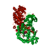
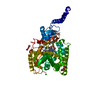
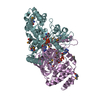
| Title | Precise Resting State of Thermus thermophilus SecYEG | ||||||
 Components Components |
| ||||||
 Keywords Keywords | PROTEIN TRANSPORT/IMMUNE SYSTEM / translocon / membrane protein / PROTEIN TRANSPORT-IMMUNE SYSTEM complex | ||||||
| Function / homology |  Function and homology information Function and homology informationprotein transport by the Sec complex / intracellular protein transmembrane transport / protein-transporting ATPase activity / transmembrane protein transporter activity / protein secretion / protein targeting / plasma membrane Similarity search - Function | ||||||
| Biological species |   Thermus thermophilus (bacteria) Thermus thermophilus (bacteria) | ||||||
| Method |  X-RAY DIFFRACTION / SYNCHROTRON / MOLECULAR REPLACEMENT / Resolution: 2.724 Å X-RAY DIFFRACTION / SYNCHROTRON / MOLECULAR REPLACEMENT / Resolution: 2.724 Å | ||||||
 Authors Authors | Tanaka, Y. / Sugano, Y. / Takemoto, M. / Kusakizako, T. / Kumazaki, K. / Ishitani, R. / Nureki, O. / Tsukazaki, T. | ||||||
 Citation Citation | Journal: Cell Rep / Year: 2015 Title: Crystal Structures of SecYEG in Lipidic Cubic Phase Elucidate a Precise Resting and a Peptide-Bound State. Authors: Tanaka, Y. / Sugano, Y. / Takemoto, M. / Mori, T. / Furukawa, A. / Kusakizako, T. / Kumazaki, K. / Kashima, A. / Ishitani, R. / Sugita, Y. / Nureki, O. / Tsukazaki, T. | ||||||
| History |
|
- Structure visualization
| Structure viewer | Molecule: MolmilJmol/JSmol |
|---|
- Downloads & links
-Download
| PDBx/mmCIF format | 5aww.cif.gz | 126.4 KB | Display | PDBx/mmCIF format |
|---|---|---|---|---|
| PDB format | pdb5aww.ent.gz | 97.2 KB | Display | PDB format |
| PDBx/mmJSON format | 5aww.json.gz | Tree view | PDBx/mmJSON format | |
| Others |  Other downloads Other downloads |
-Validation report
| Arichive directory | https://data.pdbj.org/pub/pdb/validation_reports/aw/5awwftp://data.pdbj.org/pub/pdb/validation_reports/aw/5aww | HTTPS FTP |
|---|
-Related structure data
| Related structure data | 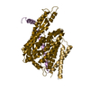 5ch4C  2zjsS C: citing same article ( S: Starting model for refinement |
|---|---|
| Similar structure data |


-Links
 PDBj
PDBj


- Assembly
| Deposited unit | 
| ||||||||
|---|---|---|---|---|---|---|---|---|---|
| 1 |
| ||||||||
| Unit cell |
|
-Components
| #1: Protein | Mass: 48957.516 Da / Num. of mol.: 1 / Mutation: L2V, R252G Source method: isolated from a genetically manipulated source Source: (gene. exp.) Thermus thermophilus (strain HB8 / ATCC 27634 / DSM 579) (bacteria)Strain: HB8 / ATCC 27634 / DSM 579 / Gene: secY, TTHA1672 Production host: References: UniProt: Q5SHQ8 | ||
|---|---|---|---|
| #2: Protein | Mass: 7072.410 Da / Num. of mol.: 1 Source method: isolated from a genetically manipulated source Source: (gene. exp.) Thermus thermophilus (strain HB8 / ATCC 27634 / DSM 579) (bacteria)Strain: HB8 / ATCC 27634 / DSM 579 / Gene: secE, TTHA0249 Production host: References: UniProt: P38383 | ||
| #3: Protein | Mass: 7943.501 Da / Num. of mol.: 1 / Fragment: UNP RESIDUES 43-116 Source method: isolated from a genetically manipulated source Source: (gene. exp.) Thermus thermophilus (strain HB8 / ATCC 27634 / DSM 579) (bacteria)Strain: HB8 / ATCC 27634 / DSM 579 / Gene: TTHA1784 Production host: References: UniProt: Q5SHE6 | ||
| #4: Chemical | ChemComp-OLC / (   Mass: 356.540 Da / Num. of mol.: 11 / Source method: obtained synthetically / Formula: C21H40O4 Mass: 356.540 Da / Num. of mol.: 11 / Source method: obtained synthetically / Formula: C21H40O4#5: Water | ChemComp-HOH / |  Mass: 18.015 Da / Num. of mol.: 7 / Source method: isolated from a natural source / Formula: H2O Mass: 18.015 Da / Num. of mol.: 7 / Source method: isolated from a natural source / Formula: H2O |
-Experimental details
-Experiment
| Experiment | Method: X-RAY DIFFRACTION |
|---|
- Sample preparation
| Crystal | Density Matthews: 3.26 Å3/Da / Density % sol: 62.25 % |
|---|---|
| Crystal grow | Temperature: 293 K / Method: lipidic cubic phase / pH: 6.5 / Details: PEG 500MME, MgSO4, Na2SO4 |
-Data collection
| Diffraction | Mean temperature: 100 K |
|---|---|
| Diffraction source | Source: SYNCHROTRON / Site: SPring-8  / Beamline: BL32XU / Wavelength: 1 Å / Beamline: BL32XU / Wavelength: 1 Å |
| Detector | Type: RAYONIX MX225HE / Detector: CCD / Date: Jan 20, 2015 |
| Radiation | Monochromator: GRAPHITE / Protocol: SINGLE WAVELENGTH / Monochromatic (M) / Laue (L): M / Scattering type: x-ray |
| Radiation wavelength | Wavelength: 1 Å / Relative weight: 1 |
| Reflection | Resolution: 2.7→50 Å / Num. obs: 21452 / % possible obs: 94.5 % / Redundancy: 4.7 % / Net I/σ(I): 10.5 |
- Processing
| Software |
| |||||||||||||||||||||||||||||||||||||||||||||||||||||||||||||||||||||||||||||||||||||||||||||||||||||||||
|---|---|---|---|---|---|---|---|---|---|---|---|---|---|---|---|---|---|---|---|---|---|---|---|---|---|---|---|---|---|---|---|---|---|---|---|---|---|---|---|---|---|---|---|---|---|---|---|---|---|---|---|---|---|---|---|---|---|---|---|---|---|---|---|---|---|---|---|---|---|---|---|---|---|---|---|---|---|---|---|---|---|---|---|---|---|---|---|---|---|---|---|---|---|---|---|---|---|---|---|---|---|---|---|---|---|---|
| Refinement | Method to determine structure: MOLECULAR REPLACEMENT Starting model: 2ZJS Resolution: 2.724→43.943 Å / SU ML: 0.4 / Cross valid method: NONE / σ(F): 1.47 / Phase error: 26.03 / Stereochemistry target values: ML
| |||||||||||||||||||||||||||||||||||||||||||||||||||||||||||||||||||||||||||||||||||||||||||||||||||||||||
| Solvent computation | Shrinkage radii: 0.9 Å / VDW probe radii: 1.11 Å / Solvent model: FLAT BULK SOLVENT MODEL | |||||||||||||||||||||||||||||||||||||||||||||||||||||||||||||||||||||||||||||||||||||||||||||||||||||||||
| Refinement step | Cycle: LAST / Resolution: 2.724→43.943 Å
| |||||||||||||||||||||||||||||||||||||||||||||||||||||||||||||||||||||||||||||||||||||||||||||||||||||||||
| Refine LS restraints |
| |||||||||||||||||||||||||||||||||||||||||||||||||||||||||||||||||||||||||||||||||||||||||||||||||||||||||
| LS refinement shell |
|
