Movie
Movie Controller


+ Open data

- Basic information
| Entry | Database: PDB / ID: 5arh | ||||||
|---|---|---|---|---|---|---|---|
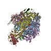
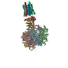
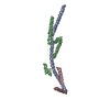
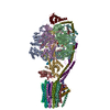
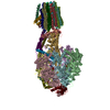
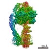
| Title | Bovine mitochondrial ATP synthase state 2a | ||||||
 Components Components |
| ||||||
 Keywords Keywords | HYDROLASE / ATP SYNTHASE / ROTARY ATPASE | ||||||
| Function / homology |  Function and homology information Function and homology informationMitochondrial protein import / Formation of ATP by chemiosmotic coupling / Cristae formation / mitochondrial proton-transporting ATP synthase complex assembly / mitochondrial envelope / proton channel activity / Mitochondrial protein degradation / proton transmembrane transporter activity / proton motive force-driven ATP synthesis / proton-transporting two-sector ATPase complex, proton-transporting domain ...Mitochondrial protein import / Formation of ATP by chemiosmotic coupling / Cristae formation / mitochondrial proton-transporting ATP synthase complex assembly / mitochondrial envelope / proton channel activity / Mitochondrial protein degradation / proton transmembrane transporter activity / proton motive force-driven ATP synthesis / proton-transporting two-sector ATPase complex, proton-transporting domain / proton motive force-driven mitochondrial ATP synthesis / H+-transporting two-sector ATPase / proton-transporting ATP synthase complex / proton-transporting ATP synthase activity, rotational mechanism / proton transmembrane transport / aerobic respiration / ADP binding / mitochondrial membrane / mitochondrial inner membrane / lipid binding / structural molecule activity / ATP hydrolysis activity / mitochondrion / ATP binding / plasma membrane Similarity search - Function | ||||||
| Biological species |  | ||||||
| Method | ELECTRON MICROSCOPY / single particle reconstruction / cryo EM / Resolution: 7.2 Å | ||||||
 Authors Authors | Zhou, A. / Rohou, A. / Schep, D.G. / Bason, J.V. / Montgomery, M.G. / Walker, J.E. / Grigorieff, N. / Rubinstein, J.L. | ||||||
 Citation Citation | Journal: Elife / Year: 2015 Title: Structure and conformational states of the bovine mitochondrial ATP synthase by cryo-EM. Authors: Anna Zhou / Alexis Rohou / Daniel G Schep / John V Bason / Martin G Montgomery / John E Walker / Nikolaus Grigorieff / John L Rubinstein /    Abstract: Adenosine triphosphate (ATP), the chemical energy currency of biology, is synthesized in eukaryotic cells primarily by the mitochondrial ATP synthase. ATP synthases operate by a rotary catalytic ...Adenosine triphosphate (ATP), the chemical energy currency of biology, is synthesized in eukaryotic cells primarily by the mitochondrial ATP synthase. ATP synthases operate by a rotary catalytic mechanism where proton translocation through the membrane-inserted FO region is coupled to ATP synthesis in the catalytic F1 region via rotation of a central rotor subcomplex. We report here single particle electron cryomicroscopy (cryo-EM) analysis of the bovine mitochondrial ATP synthase. Combining cryo-EM data with bioinformatic analysis allowed us to determine the fold of the a subunit, suggesting a proton translocation path through the FO region that involves both the a and b subunits. 3D classification of images revealed seven distinct states of the enzyme that show different modes of bending and twisting in the intact ATP synthase. Rotational fluctuations of the c8-ring within the FO region support a Brownian ratchet mechanism for proton-translocation-driven rotation in ATP synthases. | ||||||
| History |
| ||||||
| Remark 700 | SHEET DETERMINATION METHOD: DSSP THE SHEETS PRESENTED AS "AA" IN EACH CHAIN ON SHEET RECORDS BELOW ... SHEET DETERMINATION METHOD: DSSP THE SHEETS PRESENTED AS "AA" IN EACH CHAIN ON SHEET RECORDS BELOW IS ACTUALLY AN 6-STRANDED BARREL THIS IS REPRESENTED BY A 7-STRANDED SHEET IN WHICH THE FIRST AND LAST STRANDS ARE IDENTICAL. SHEET DETERMINATION METHOD: DSSP THE SHEETS PRESENTED AS "BA" IN EACH CHAIN ON SHEET RECORDS BELOW IS ACTUALLY AN 10-STRANDED BARREL THIS IS REPRESENTED BY A 11-STRANDED SHEET IN WHICH THE FIRST AND LAST STRANDS ARE IDENTICAL. SHEET DETERMINATION METHOD: DSSP THE SHEETS PRESENTED AS "CA" IN EACH CHAIN ON SHEET RECORDS BELOW IS ACTUALLY AN 10-STRANDED BARREL THIS IS REPRESENTED BY A 11-STRANDED SHEET IN WHICH THE FIRST AND LAST STRANDS ARE IDENTICAL. SHEET DETERMINATION METHOD: DSSP THE SHEETS PRESENTED AS "EA" IN EACH CHAIN ON SHEET RECORDS BELOW IS ACTUALLY AN 6-STRANDED BARREL THIS IS REPRESENTED BY A 7-STRANDED SHEET IN WHICH THE FIRST AND LAST STRANDS ARE IDENTICAL. |
- Structure visualization
| Movie |
Movie viewer |
|---|---|
| Structure viewer | Molecule: MolmilJmol/JSmol |
- Downloads & links
-Download
| PDBx/mmCIF format | 5arh.cif.gz | 564.9 KB | Display | PDBx/mmCIF format |
|---|---|---|---|---|
| PDB format | pdb5arh.ent.gz | 350.3 KB | Display | PDB format |
| PDBx/mmJSON format | 5arh.json.gz | Tree view | PDBx/mmJSON format | |
| Others |  Other downloads Other downloads |
-Validation report
| Summary document | 5arh_validation.pdf.gz | 856 KB | Display | wwPDB validaton report |
|---|---|---|---|---|
| Full document | 5arh_full_validation.pdf.gz | 896.7 KB | Display | |
| Data in XML | 5arh_validation.xml.gz | 90.5 KB | Display | |
| Data in CIF | 5arh_validation.cif.gz | 160.4 KB | Display | |
| Arichive directory | https://data.pdbj.org/pub/pdb/validation_reports/ar/5arhftp://data.pdbj.org/pub/pdb/validation_reports/ar/5arh | HTTPS FTP |
-Related structure data
| Related structure data | 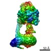 3166MC 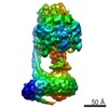 3164C 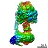 3165C 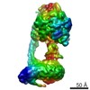 3167C 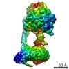 3168C 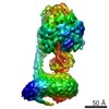 3169C 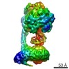 3170C 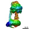 3181C 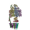 5araC  5areC 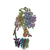 5ariC  5fijC  5fikC 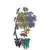 5filC C: citing same article ( M: map data used to model this data |
|---|---|
| Similar structure data |


-Links
 PDBj
PDBj


- Assembly
| Deposited unit | 
|
|---|---|
| 1 |
|
-Components
-ATP SYNTHASE SUBUNIT ... , 8 types, 12 molecules ABCDEFGHISUW
| #1: Protein | Mass: 55301.207 Da / Num. of mol.: 3 / Fragment: UNP RESIDUES 44-553 / Source method: isolated from a natural source / Source: (natural) #2: Protein | Mass: 51757.836 Da / Num. of mol.: 3 / Fragment: UNP RESIDUES 47-528 / Source method: isolated from a natural source / Source: (natural) References: UniProt: P00829, H+-transporting two-sector ATPase #3: Protein | | Mass: 30300.760 Da / Num. of mol.: 1 / Fragment: UNP RESIDUES 26-298 / Source method: isolated from a natural source / Source: (natural) #4: Protein | | Mass: 15074.813 Da / Num. of mol.: 1 / Fragment: UNP RESIDUES 23-168 / Source method: isolated from a natural source / Source: (natural) #5: Protein/peptide | | Mass: 5662.693 Da / Num. of mol.: 1 / Fragment: UNP RESIDUES 2-51 / Source method: isolated from a natural source / Source: (natural) #7: Protein | | Mass: 20989.803 Da / Num. of mol.: 1 / Fragment: UNP RESIDUES 24-213 Source method: isolated from a genetically manipulated source Source: (gene. exp.)  #9: Protein | | Mass: 14167.169 Da / Num. of mol.: 1 / Fragment: UNP RESIDUES 2-125 Source method: isolated from a genetically manipulated source Source: (gene. exp.) #11: Protein | | Mass: 23717.578 Da / Num. of mol.: 1 / Fragment: UNP RESIDUES 10-226 / Source method: isolated from a natural source / Source: (natural) |
|---|
-ATP SYNTHASE F(0) COMPLEX SUBUNIT ... , 2 types, 9 molecules JKLMNOPQT
| #6: Protein | Mass: 7293.593 Da / Num. of mol.: 8 / Fragment: UNP RESIDUES 63-134 / Source method: isolated from a natural source / Source: (natural) #8: Protein | | Mass: 20335.625 Da / Num. of mol.: 1 / Fragment: UNP RESIDUES 76-249 Source method: isolated from a genetically manipulated source Source: (gene. exp.) |
|---|
-Protein , 1 types, 1 molecules V
| #10: Protein | Mass: 9118.253 Da / Num. of mol.: 1 / Fragment: UNP RESIDUES 32-108 Source method: isolated from a genetically manipulated source Source: (gene. exp.) |
|---|
-Experimental details
-Experiment
| Experiment | Method: ELECTRON MICROSCOPY |
|---|---|
| EM experiment | Aggregation state: PARTICLE / 3D reconstruction method: single particle reconstruction |
- Sample preparation
| Component | Name: BOVINE MITOCHONDRIAL ATP SYNTHASE / Type: COMPLEX |
|---|---|
| Buffer solution | Name: 20 MM TRIS-HCL, 100 MM NACL, 10% (V/V) DODECYLMALTOSIDE, 2 MM ATP, 0.02% (WT/V) NAN3 pH: 7.2 Details: 20 MM TRIS-HCL, 100 MM NACL, 10% (V/V) DODECYLMALTOSIDE, 2 MM ATP, 0.02% (WT/V) NAN3 |
| Specimen | Conc.: 8 mg/ml / Embedding applied: NO / Shadowing applied: NO / Staining applied: NO / Vitrification applied: YES |
| Specimen support | Details: HOLEY CARBON |
| Vitrification | Instrument: FEI VITROBOT MARK III / Cryogen name: ETHANE-PROPANE Details: VITRIFICATION 1 -- CRYOGEN- ETHANE-PROPANE MIXTURE, HUMIDITY- 100, INSTRUMENT- FEI VITROBOT MARK III, METHOD- BLOT FOR 27 SECONDS BEFORE PLUNGING, |
- Electron microscopy imaging
| Experimental equipment |  Model: Titan Krios / Image courtesy: FEI Company |
|---|---|
| Microscopy | Model: FEI TITAN KRIOS / Date: Mar 15, 2015 Details: K2 SUMMIT DIRECT DETECTOR DEVICE (GATAN INC.) OPERATED IN SUPER-RESOLUTION MODE WITH A 1.64 ANGSTROM PHYSICAL PIXEL AND 0. 82 ANGSTROM SUPER- RESOLUTION PIXEL. WITH NO SPECIMEN PRESENT, THE ...Details: K2 SUMMIT DIRECT DETECTOR DEVICE (GATAN INC.) OPERATED IN SUPER-RESOLUTION MODE WITH A 1.64 ANGSTROM PHYSICAL PIXEL AND 0. 82 ANGSTROM SUPER- RESOLUTION PIXEL. WITH NO SPECIMEN PRESENT, THE RATE OF EXPOSURE OF THE DETECTOR WAS 8 ELECTRONS PER PIXEL PER SECOND. EXPOSURE- FRACTIONATED MOVIES OF 20.1 S WERE RECORDED AS STACKS OF 67 FRAMES, SO THAT SELECTED SPECIMEN AREAS WERE EXPOSED WITH A TOTAL OF 60.3 ELECTRONS PER SQUARE ANGSTROM. |
| Electron gun | Electron source:  FIELD EMISSION GUN / Accelerating voltage: 300 kV / Illumination mode: FLOOD BEAM FIELD EMISSION GUN / Accelerating voltage: 300 kV / Illumination mode: FLOOD BEAM |
| Electron lens | Mode: BRIGHT FIELD / Nominal magnification: 18000 X / Calibrated magnification: 30487 X / Nominal defocus max: 4100 nm / Nominal defocus min: 1200 nm / Cs: 2.7 mm |
| Specimen holder | Temperature: 80 K |
| Image recording | Electron dose: 60.3 e/Å2 / Film or detector model: GATAN K2 (4k x 4k) |
- Processing
| EM software |
| ||||||||||||||||||||||||||||
|---|---|---|---|---|---|---|---|---|---|---|---|---|---|---|---|---|---|---|---|---|---|---|---|---|---|---|---|---|---|
| CTF correction | Details: EACH PARTICLE | ||||||||||||||||||||||||||||
| Symmetry | Point symmetry: C1 (asymmetric) | ||||||||||||||||||||||||||||
| 3D reconstruction | Method: PROJECTION MATCHING AND MAXIMUM LIKELIHOOD CLASSIFICATION Resolution: 7.2 Å / Num. of particles: 19250 Details: THE A SUBUNIT WAS MODELLED USING EVOLUTIONARY CO-VARIANCE. THE N-TERMINAL TRANSMEMBRANE HELICES OF THE B SUBUNIT WERE MODELLED BASED ON TRANSMEMBRANE HELIX PREDICTIONS. SUBMISSION BASED ON ...Details: THE A SUBUNIT WAS MODELLED USING EVOLUTIONARY CO-VARIANCE. THE N-TERMINAL TRANSMEMBRANE HELICES OF THE B SUBUNIT WERE MODELLED BASED ON TRANSMEMBRANE HELIX PREDICTIONS. SUBMISSION BASED ON EXPERIMENTAL DATA FROM EMDB EMD-3166. (DEPOSITION ID: 13798). Symmetry type: POINT | ||||||||||||||||||||||||||||
| Atomic model building | Protocol: FLEXIBLE FIT / Details: METHOD--FLEXIBLE FITTING | ||||||||||||||||||||||||||||
| Atomic model building |
| ||||||||||||||||||||||||||||
| Refinement | Highest resolution: 7.2 Å | ||||||||||||||||||||||||||||
| Refinement step | Cycle: LAST / Highest resolution: 7.2 Å
|