 Movie
Movie Controller


+ Open data

- Basic information
| Entry | Database: PDB / ID: 5ao5 | ||||||
|---|---|---|---|---|---|---|---|
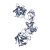
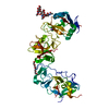
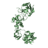
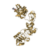
| Title | Endo180 D1-4, monoclinic form | ||||||
 Components Components | C-TYPE MANNOSE RECEPTOR 2 | ||||||
 Keywords Keywords | ENDOCYTOSIS / ENDOCYTIC RECEPTOR / FIBRONECTIN TYPE II DOMAIN / C-TYPE LECTIN DOMAIN / COLLAGEN / GELATIN | ||||||
| Function / homology |  Function and homology information Function and homology informationCross-presentation of soluble exogenous antigens (endosomes) / collagen catabolic process / collagen binding / endocytosis / osteoblast differentiation / carbohydrate binding / signaling receptor activity / focal adhesion / membrane Similarity search - Function | ||||||
| Biological species |  HOMO SAPIENS (human) HOMO SAPIENS (human) | ||||||
| Method |  X-RAY DIFFRACTION / SYNCHROTRON / MOLECULAR REPLACEMENT / Resolution: 2.48 Å X-RAY DIFFRACTION / SYNCHROTRON / MOLECULAR REPLACEMENT / Resolution: 2.48 Å | ||||||
 Authors Authors | Paracuellos, P. / Briggs, D.C. / Carafoli, F. / Loncar, T. / Hohenester, E. | ||||||
 Citation Citation | Journal: Structure / Year: 2015 Title: Insights Into Collagen Uptake by C-Type Mannose Receptors from the Crystal Structure of Endo180 Domains 1-4. Authors: Paracuellos, P. / Briggs, D.C. / Carafoli, F. / Loncar, T. / Hohenester, E. | ||||||
| History |
|
- Structure visualization
| Structure viewer | Molecule: MolmilJmol/JSmol |
|---|
- Downloads & links
-Download
| PDBx/mmCIF format | 5ao5.cif.gz | 329.7 KB | Display | PDBx/mmCIF format |
|---|---|---|---|---|
| PDB format | pdb5ao5.ent.gz | 269.5 KB | Display | PDB format |
| PDBx/mmJSON format | 5ao5.json.gz | Tree view | PDBx/mmJSON format | |
| Others |  Other downloads Other downloads |
-Validation report
| Arichive directory | https://data.pdbj.org/pub/pdb/validation_reports/ao/5ao5ftp://data.pdbj.org/pub/pdb/validation_reports/ao/5ao5 | HTTPS FTP |
|---|
-Related structure data
| Related structure data | 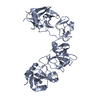 5ao6C 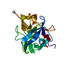 1dqgS 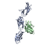 1tdqS  3m7pS C: citing same article ( S: Starting model for refinement |
|---|---|
| Similar structure data |


-Links
 PDBj
PDBj


- Assembly
| Deposited unit | 
| ||||||||
|---|---|---|---|---|---|---|---|---|---|
| 1 | 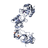
| ||||||||
| 2 | 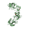
| ||||||||
| Unit cell |
|
-Components
| #1: Protein | Mass: 54592.484 Da / Num. of mol.: 2 / Fragment: DOMAINS 1-4, RESIDUES 35-511 Source method: isolated from a genetically manipulated source Source: (gene. exp.) HOMO SAPIENS (human) / Plasmid: PCEP-PU / Cell line (production host): HEK293 / Production host: HOMO SAPIENS (human) / References: UniProt: Q9UBG0#2: Chemical | ChemComp-NA /   Mass: 22.990 Da / Num. of mol.: 4 / Source method: obtained synthetically / Formula: Na Mass: 22.990 Da / Num. of mol.: 4 / Source method: obtained synthetically / Formula: Na#3: Chemical | ChemComp-SO4 /   Mass: 96.063 Da / Num. of mol.: 4 / Source method: obtained synthetically / Formula: SO4 Mass: 96.063 Da / Num. of mol.: 4 / Source method: obtained synthetically / Formula: SO4#4: Water | ChemComp-HOH / |  Mass: 18.015 Da / Num. of mol.: 163 / Source method: isolated from a natural source / Formula: H2O Mass: 18.015 Da / Num. of mol.: 163 / Source method: isolated from a natural source / Formula: H2OHas protein modification | Y | Sequence details | VECTOR-DERIVED APLA AT N-TERMINUS. | |
|---|
-Experimental details
-Experiment
| Experiment | Method: X-RAY DIFFRACTION / Number of used crystals: 1 |
|---|
- Sample preparation
| Crystal | Density Matthews: 3.36 Å3/Da / Density % sol: 62 % / Description: NONE |
|---|---|
| Crystal grow | pH: 7.5 / Details: pH 7.5 |
-Data collection
| Diffraction | Mean temperature: 100 K |
|---|---|
| Diffraction source | Source: SYNCHROTRON / Site: Diamond  / Beamline: I04-1 / Wavelength: 0.92 / Beamline: I04-1 / Wavelength: 0.92 |
| Detector | Type: DECTRIS PIXEL / Detector: PIXEL / Date: Oct 5, 2014 |
| Radiation | Protocol: SINGLE WAVELENGTH / Monochromatic (M) / Laue (L): M / Scattering type: x-ray |
| Radiation wavelength | Wavelength: 0.92 Å / Relative weight: 1 |
| Reflection | Resolution: 2.48→62.8 Å / Num. obs: 50153 / % possible obs: 97.8 % / Observed criterion σ(I): 0 / Redundancy: 3.3 % / Biso Wilson estimate: 46.16 Å2 / Rmerge(I) obs: 0.05 / Net I/σ(I): 15.4 |
| Reflection shell | Resolution: 2.48→2.54 Å / Redundancy: 3.4 % / Rmerge(I) obs: 0.48 / Mean I/σ(I) obs: 2.2 / % possible all: 98.4 |
- Processing
| Software |
| |||||||||||||||||||||||||||||||||||||||||||||||||||||||||||||||||||||||||||||||||||||||||||||||||||||||||||||||||||||||||||||||||||||
|---|---|---|---|---|---|---|---|---|---|---|---|---|---|---|---|---|---|---|---|---|---|---|---|---|---|---|---|---|---|---|---|---|---|---|---|---|---|---|---|---|---|---|---|---|---|---|---|---|---|---|---|---|---|---|---|---|---|---|---|---|---|---|---|---|---|---|---|---|---|---|---|---|---|---|---|---|---|---|---|---|---|---|---|---|---|---|---|---|---|---|---|---|---|---|---|---|---|---|---|---|---|---|---|---|---|---|---|---|---|---|---|---|---|---|---|---|---|---|---|---|---|---|---|---|---|---|---|---|---|---|---|---|---|---|
| Refinement | Method to determine structure: MOLECULAR REPLACEMENT Starting model: PDB ENTRIES 1TDQ, 3M7P, 1DQG Resolution: 2.48→60.158 Å / SU ML: 0.33 / σ(F): 1.34 / Phase error: 28.32 / Stereochemistry target values: ML
| |||||||||||||||||||||||||||||||||||||||||||||||||||||||||||||||||||||||||||||||||||||||||||||||||||||||||||||||||||||||||||||||||||||
| Solvent computation | Shrinkage radii: 0.9 Å / VDW probe radii: 1.11 Å / Solvent model: FLAT BULK SOLVENT MODEL | |||||||||||||||||||||||||||||||||||||||||||||||||||||||||||||||||||||||||||||||||||||||||||||||||||||||||||||||||||||||||||||||||||||
| Refinement step | Cycle: LAST / Resolution: 2.48→60.158 Å
| |||||||||||||||||||||||||||||||||||||||||||||||||||||||||||||||||||||||||||||||||||||||||||||||||||||||||||||||||||||||||||||||||||||
| Refine LS restraints |
| |||||||||||||||||||||||||||||||||||||||||||||||||||||||||||||||||||||||||||||||||||||||||||||||||||||||||||||||||||||||||||||||||||||
| LS refinement shell |
|
