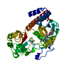
Entry Database : PDB / ID : 5ajgTitle Structure of Infrared Fluorescent Protein IFP1.4 AT 1.11 Angstrom resolution BACTERIOPHYTOCHROME Keywords Function / homology Function Domain/homology Component
/ / / / / / / / / / / / / / / / / / / / / / / / / / / / / / / / / / / / / / / / / / / Biological species DEINOCOCCUS RADIODURANS (radioresistant)Method / / / Resolution : 1.11 Å Authors Lafaye, C. / Shu, X. / Royant, A. Journal : Biochemistry / Year : 2016Title : Structural Determinants of Improved Fluorescence in a Family of Bacteriophytochrome-Based Infrared Fluorescent Proteins: Insights from Continuum Electrostatic Calculations and Molecular Dynamics Simulations.Authors : Feliks, M. / Lafaye, C. / Shu, X. / Royant, A. / Field, M. History Deposition Feb 24, 2015 Deposition site / Processing site Revision 1.0 Mar 9, 2016 Provider / Type Revision 1.1 Aug 10, 2016 Group Revision 1.2 Aug 17, 2016 Group Revision 1.3 Aug 24, 2016 Group Revision 1.4 Jan 10, 2024 Group Data collection / Database references ... Data collection / Database references / Derived calculations / Other / Refinement description / Structure summary Category chem_comp / chem_comp_atom ... chem_comp / chem_comp_atom / chem_comp_bond / database_2 / entity / pdbx_database_status / pdbx_entity_nonpoly / pdbx_initial_refinement_model / struct_conn / struct_site Item _chem_comp.name / _database_2.pdbx_DOI ... _chem_comp.name / _database_2.pdbx_DOI / _database_2.pdbx_database_accession / _entity.pdbx_description / _pdbx_database_status.status_code_sf / _pdbx_entity_nonpoly.name / _struct_conn.pdbx_leaving_atom_flag / _struct_site.pdbx_auth_asym_id / _struct_site.pdbx_auth_comp_id / _struct_site.pdbx_auth_seq_id Revision 1.5 Nov 20, 2024 Group / Category / pdbx_modification_feature
Show all Show less
 Movie
Movie Controller

 Yorodumi
 Open data
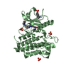
 Basic information
 Components
 Keywords
 Function and homology information
 DEINOCOCCUS RADIODURANS (radioresistant)
 X-RAY DIFFRACTION /
  Authors
 Citation
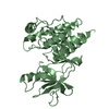
 Structure visualization
 Downloads & links
 Other downloads
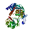


 PDBj
PDBj


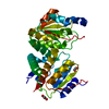
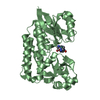
 Assembly
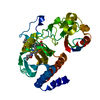
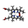
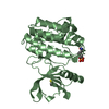
 Mass: 585.670 Da / Num. of mol.: 1 / Source method: obtained synthetically / Formula: C33H37N4O6
 Mass: 18.015 Da / Num. of mol.: 262 / Source method: isolated from a natural source / Formula: H2O
 Sample preparation
 / Beamline: ID29 / Wavelength: 0.976
  Processing