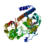
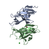
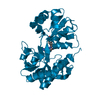
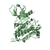
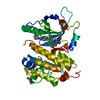
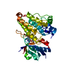
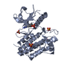
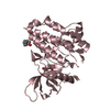
Entry Database : PDB / ID : 4cqhTitle Structure of Infrared Fluorescent Protein IFP2.0 BACTERIOPHYTOCHROME Keywords / Function / homology Function Domain/homology Component
/ / / / / / / / / / / / / / / / / / / / / / / / / / / / / / / / / / / / / / / / / / / Biological species DEINOCOCCUS RADIODURANS (radioresistant)Method / / / Resolution : 1.14 Å Authors Lafaye, C. / Yu, D. / Noirclerc-Savoye, M. / Shu, X. / Royant, A. Journal : Nat.Commun. / Year : 2014Title : An Improved Monomeric Infrared Fluorescent Protein for Neuronal and Tumour Brain Imaging.
Authors :
Yu, D. / Gustafson, W.C. / Han, C. / Lafaye, C. / Noirclerc-Savoye, M. / Ge, W. / Thayer, D.A. / Huang, H. / Kornberg, T.B. / Royant, A. / Jan, L.Y. / Jan, Y.N. / Weiss, W.A. / Shu, X. History Deposition Feb 17, 2014 Deposition site / Processing site Revision 1.0 May 28, 2014 Provider / Type Revision 1.1 Dec 20, 2023 Group Data collection / Database references ... Data collection / Database references / Derived calculations / Other / Refinement description / Structure summary Category chem_comp / chem_comp_atom ... chem_comp / chem_comp_atom / chem_comp_bond / database_2 / entity / pdbx_database_status / pdbx_entity_nonpoly / pdbx_initial_refinement_model / pdbx_struct_conn_angle / struct_conn / struct_site Item _chem_comp.name / _database_2.pdbx_DOI ... _chem_comp.name / _database_2.pdbx_DOI / _database_2.pdbx_database_accession / _entity.pdbx_description / _pdbx_database_status.status_code_sf / _pdbx_entity_nonpoly.name / _pdbx_struct_conn_angle.ptnr1_auth_seq_id / _pdbx_struct_conn_angle.ptnr3_auth_seq_id / _pdbx_struct_conn_angle.value / _struct_conn.pdbx_dist_value / _struct_conn.pdbx_leaving_atom_flag / _struct_conn.ptnr2_auth_seq_id / _struct_site.pdbx_auth_asym_id / _struct_site.pdbx_auth_comp_id / _struct_site.pdbx_auth_seq_id Revision 1.2 Nov 6, 2024 Group / Category / pdbx_modification_feature / Item
Show all Show less
 Movie
Movie Controller


 Open data
 Basic information
 Components
 Keywords
 Function and homology information
 DEINOCOCCUS RADIODURANS (radioresistant)
 X-RAY DIFFRACTION /
  Authors
 Citation
 Structure visualization
 Downloads & links
 Other downloads
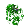


 PDBj
PDBj


 Assembly
 Mass: 585.670 Da / Num. of mol.: 1 / Source method: obtained synthetically / Formula: C33H37N4O6
 Mass: 22.990 Da / Num. of mol.: 1 / Source method: obtained synthetically / Formula: Na
 Mass: 18.015 Da / Num. of mol.: 291 / Source method: isolated from a natural source / Formula: H2O
 Sample preparation
 / Beamline: ID23-2 / Wavelength: 0.873
  Processing