 Movie
Movie Controller


+ Open data

- Basic information
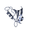
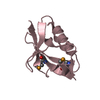
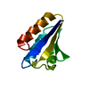
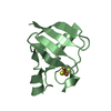
| Entry | Database: PDB / ID: 5a7l | ||||||
|---|---|---|---|---|---|---|---|
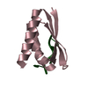
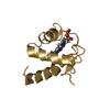
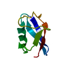
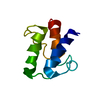
| Title | TP901-1 CI NTD (res 1-80) | ||||||
 Components Components | CI | ||||||
 Keywords Keywords | TRANSCRIPTION | ||||||
| Function / homology |  Function and homology information Function and homology information | ||||||
| Biological species |  LACTOCOCCUS PHAGE (virus) LACTOCOCCUS PHAGE (virus) | ||||||
| Method |  X-RAY DIFFRACTION / SYNCHROTRON / MOLECULAR REPLACEMENT / Resolution: 2.103 Å X-RAY DIFFRACTION / SYNCHROTRON / MOLECULAR REPLACEMENT / Resolution: 2.103 Å | ||||||
 Authors Authors | Frandsen, K.E.H. / Rasmussen, K.K. / Lo Leggio, L. | ||||||
 Citation Citation | Journal: Sci.Rep. / Year: 2016 Title: Structural and Dynamics Studies of a Truncated Variant of Ci Repressor from Bacteriophage Tp901-1. Authors: Rasmussen, K.K. / Frandsen, K.E.H. / Boeri Erba, E. / Pedersen, M. / Varming, A.K. / Hammer, K. / Kilstrup, M. / Thulstrup, P.W. / Blackledge, M. / Jensen, M.R. / Lo Leggio, L. | ||||||
| History |
|
- Structure visualization
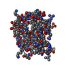
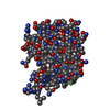
| Structure viewer | Molecule: MolmilJmol/JSmol |
|---|
- Downloads & links
-Download
| PDBx/mmCIF format | 5a7l.cif.gz | 40.8 KB | Display | PDBx/mmCIF format |
|---|---|---|---|---|
| PDB format | pdb5a7l.ent.gz | 29.3 KB | Display | PDB format |
| PDBx/mmJSON format | 5a7l.json.gz | Tree view | PDBx/mmJSON format | |
| Others |  Other downloads Other downloads |
-Validation report
| Arichive directory | https://data.pdbj.org/pub/pdb/validation_reports/a7/5a7lftp://data.pdbj.org/pub/pdb/validation_reports/a7/5a7l | HTTPS FTP |
|---|
-Related structure data
| Related structure data |  3zhiS S: Starting model for refinement |
|---|---|
| Similar structure data |
-Links
 PDBj
PDBj


- Assembly
| Deposited unit | 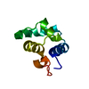
| ||||||||
|---|---|---|---|---|---|---|---|---|---|
| 1 |
| ||||||||
| Unit cell |
|
-Components
| #1: Protein | Mass: 9127.633 Da / Num. of mol.: 1 / Fragment: UNP RESIDUES 1-80 / Mutation: YES Source method: isolated from a genetically manipulated source Source: (gene. exp.) LACTOCOCCUS PHAGE (virus) / Strain: TP901-1 / Production host:  |
|---|---|
| #2: Water | ChemComp-HOH /  Mass: 18.015 Da / Num. of mol.: 39 / Source method: isolated from a natural source / Formula: H2O Mass: 18.015 Da / Num. of mol.: 39 / Source method: isolated from a natural source / Formula: H2O |
-Experimental details
-Experiment
| Experiment | Method: X-RAY DIFFRACTION |
|---|
- Sample preparation
| Crystal | Density Matthews: 2.06 Å3/Da / Density % sol: 40.39 % / Description: NONE |
|---|
-Data collection
| Diffraction | Mean temperature: 100 K |
|---|---|
| Diffraction source | Source: SYNCHROTRON / Site: PETRA III, DESY  / Beamline: P11 / Wavelength: 1.033 / Beamline: P11 / Wavelength: 1.033 |
| Detector | Type: DECTRIS PILATUS 6M / Detector: PIXEL |
| Radiation | Protocol: SINGLE WAVELENGTH / Monochromatic (M) / Laue (L): M / Scattering type: x-ray |
| Radiation wavelength | Wavelength: 1.033 Å / Relative weight: 1 |
| Reflection | Resolution: 2.1→30 Å / Num. obs: 4277 / % possible obs: 90.6 % / Observed criterion σ(I): -3 / Redundancy: 3.52 % / Biso Wilson estimate: 31.4 Å2 / Rmerge(I) obs: 0.17 / Net I/σ(I): 5.68 |
| Reflection shell | Resolution: 2.1→2.16 Å / Redundancy: 2.89 % / Rmerge(I) obs: 0.81 / Mean I/σ(I) obs: 1.73 / % possible all: 87.3 |
- Processing
| Software |
| ||||||||||||||||||||||||||||
|---|---|---|---|---|---|---|---|---|---|---|---|---|---|---|---|---|---|---|---|---|---|---|---|---|---|---|---|---|---|
| Refinement | Method to determine structure: MOLECULAR REPLACEMENT Starting model: PDB ENTRY 3ZHI Resolution: 2.103→29.911 Å / SU ML: 0.26 / σ(F): 1.37 / Phase error: 26.65 / Stereochemistry target values: ML
| ||||||||||||||||||||||||||||
| Solvent computation | Shrinkage radii: 0.9 Å / VDW probe radii: 1.11 Å / Solvent model: FLAT BULK SOLVENT MODEL | ||||||||||||||||||||||||||||
| Refinement step | Cycle: LAST / Resolution: 2.103→29.911 Å
| ||||||||||||||||||||||||||||
| Refine LS restraints |
| ||||||||||||||||||||||||||||
| LS refinement shell |
|
