 Movie
Movie Controller

[English] 日本語
 Yorodumi
- PDB-5a7f: Comparison of the structure and activity of glycosylated and agly... -
+ Open data

- Basic information
| Entry | Database: PDB / ID: 5a7f | ||||||
|---|---|---|---|---|---|---|---|
| Title | Comparison of the structure and activity of glycosylated and aglycosylated Human Carboxylesterase 1 | ||||||
 Components Components | LIVER CARBOXYLESTERASE 1 | ||||||
 Keywords Keywords | HYDROLASE / ESTERASE | ||||||
| Function / homology |  Function and homology information Function and homology informationcholesterol ester hydrolysis involved in cholesterol transport / methylumbelliferyl-acetate deacetylase / methylumbelliferyl-acetate deacetylase activity / regulation of bile acid secretion / sterol esterase / sterol ester esterase activity / medium-chain fatty acid metabolic process / carboxylesterase / Physiological factors / carboxylesterase activity ...cholesterol ester hydrolysis involved in cholesterol transport / methylumbelliferyl-acetate deacetylase / methylumbelliferyl-acetate deacetylase activity / regulation of bile acid secretion / sterol esterase / sterol ester esterase activity / medium-chain fatty acid metabolic process / carboxylesterase / Physiological factors / carboxylesterase activity / regulation of bile acid biosynthetic process / cellular response to cholesterol / reverse cholesterol transport / positive regulation of cholesterol metabolic process / Phase I - Functionalization of compounds / carboxylic ester hydrolase activity / cholesterol biosynthetic process / Aspirin ADME / negative regulation of cholesterol storage / positive regulation of cholesterol efflux / cellular response to low-density lipoprotein particle stimulus / cholesterol metabolic process / Metabolism of Angiotensinogen to Angiotensins / lipid catabolic process / epithelial cell differentiation / lipid droplet / cholesterol homeostasis / response to toxic substance / endoplasmic reticulum lumen / endoplasmic reticulum / cytoplasm / cytosol Similarity search - Function | ||||||
| Biological species |  HOMO SAPIENS (human) HOMO SAPIENS (human) | ||||||
| Method |  X-RAY DIFFRACTION / SYNCHROTRON / MOLECULAR REPLACEMENT / Resolution: 1.86 Å X-RAY DIFFRACTION / SYNCHROTRON / MOLECULAR REPLACEMENT / Resolution: 1.86 Å | ||||||
 Authors Authors | Arena de Souza, V. / Scott, D.J. / Charlton, M. / Walsh, M.A. / Owen, R.J. | ||||||
 Citation Citation | Journal: Plos One / Year: 2015 Title: Comparison of the Structure and Activity of Glycosylated and Aglycosylated Human Carboxylesterase 1. Authors: Arena De Souza, V. / Scott, D.J. / Nettleship, J.E. / Rahman, N. / Charlton, M.H. / Walsh, M.A. / Owens, R.J. | ||||||
| History |
|
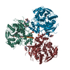
- Structure visualization
| Structure viewer | Molecule: MolmilJmol/JSmol |
|---|
- Downloads & links
-Download
| PDBx/mmCIF format | 5a7f.cif.gz | 220.5 KB | Display | PDBx/mmCIF format |
|---|---|---|---|---|
| PDB format | pdb5a7f.ent.gz | 177.3 KB | Display | PDB format |
| PDBx/mmJSON format | 5a7f.json.gz | Tree view | PDBx/mmJSON format | |
| Others |  Other downloads Other downloads |
-Validation report
| Arichive directory | https://data.pdbj.org/pub/pdb/validation_reports/a7/5a7fftp://data.pdbj.org/pub/pdb/validation_reports/a7/5a7f | HTTPS FTP |
|---|
-Related structure data
| Related structure data | 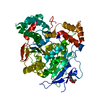 5a7gC 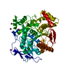 5a7hC 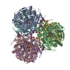 2h7cS C: citing same article ( S: Starting model for refinement |
|---|---|
| Similar structure data |

-Links
 PDBj
PDBj


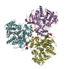
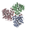
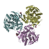
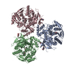
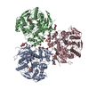
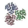
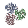
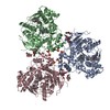
- Assembly
| Deposited unit | 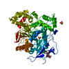
| ||||||||
|---|---|---|---|---|---|---|---|---|---|
| 1 | 
| ||||||||
| Unit cell |
|
-Components
| #1: Protein | Mass: 58672.227 Da / Num. of mol.: 1 / Fragment: UNP RESIDUES 21-553 Source method: isolated from a genetically manipulated source Source: (gene. exp.) HOMO SAPIENS (human) / Cell line: HEK 323 / Organ: LIVER / Plasmid: POPINTTG / Cell line (production host): HEK 323 / Production host: HOMO SAPIENS (human)References: UniProt: P23141-3, UniProt: P23141*PLUS, carboxylesterase, carboxylesterase, methylumbelliferyl-acetate deacetylase | ||||||
|---|---|---|---|---|---|---|---|
| #2: Sugar | ChemComp-NAG /   Type: D-saccharide, beta linking / Mass: 221.208 Da / Num. of mol.: 1 Type: D-saccharide, beta linking / Mass: 221.208 Da / Num. of mol.: 1Source method: isolated from a genetically manipulated source Formula: C8H15NO6 | ||||||
| #3: Chemical |   Mass: 94.971 Da / Num. of mol.: 2 / Source method: obtained synthetically / Formula: PO4 Mass: 94.971 Da / Num. of mol.: 2 / Source method: obtained synthetically / Formula: PO4#4: Water | ChemComp-HOH / |  Mass: 18.015 Da / Num. of mol.: 270 / Source method: isolated from a natural source / Formula: H2O Mass: 18.015 Da / Num. of mol.: 270 / Source method: isolated from a natural source / Formula: H2OHas protein modification | Y | Nonpolymer details | N-ACETYL-D-GLUCOSAMIN | |
-Experimental details
-Experiment
| Experiment | Method: X-RAY DIFFRACTION / Number of used crystals: 1 |
|---|
- Sample preparation
| Crystal | Density Matthews: 2.5 Å3/Da / Density % sol: 50.8 % / Description: NONE |
|---|---|
| Crystal grow | Temperature: 293 K / pH: 6.5 Details: CRYSTALS WERE GROWN AT 20C IN 0.1 M MES/IMIDAZOLE, PH 6.5 CONTAINING 0.03 M EACH OF DIETHYLENE GLYCOL, TRIETHYLENE GLYCOL, TETRAETHYLENE GLYCOL, PENTAETHYLENE GLYCOL, 10 % (W/V) POLYETHYLENE ...Details: CRYSTALS WERE GROWN AT 20C IN 0.1 M MES/IMIDAZOLE, PH 6.5 CONTAINING 0.03 M EACH OF DIETHYLENE GLYCOL, TRIETHYLENE GLYCOL, TETRAETHYLENE GLYCOL, PENTAETHYLENE GLYCOL, 10 % (W/V) POLYETHYLENE GLYCOL 4000, 20% (W/V) GLYCEROL |
-Data collection
| Diffraction | Mean temperature: 100 K |
|---|---|
| Diffraction source | Source: SYNCHROTRON / Site: Diamond  / Beamline: I04 / Wavelength: 0.9795 / Beamline: I04 / Wavelength: 0.9795 |
| Detector | Type: ADSC QUANTUM 315 / Detector: CCD / Date: Oct 10, 2011 / Details: MIRRORS |
| Radiation | Monochromator: SI111 / Protocol: SINGLE WAVELENGTH / Monochromatic (M) / Laue (L): M / Scattering type: x-ray |
| Radiation wavelength | Wavelength: 0.9795 Å / Relative weight: 1 |
| Reflection | Resolution: 1.86→50 Å / Num. obs: 46879 / % possible obs: 97 % / Observed criterion σ(I): -3 / Redundancy: 2.8 % / Rmerge(I) obs: 0.06 / Net I/σ(I): 17.4 |
| Reflection shell | Resolution: 1.86→1.88 Å / Redundancy: 2.3 % / Rmerge(I) obs: 0.43 / Mean I/σ(I) obs: 1.8 / % possible all: 71.5 |
- Processing
| Software |
| ||||||||||||||||||||||||||||||||||||||||||||||||||||||||||||||||||||||||||||||||||||||||||||||||||||||||||||||||||||||||||||||||||||||||||||||||||||||||||||||||||||||||||||||||||||||
|---|---|---|---|---|---|---|---|---|---|---|---|---|---|---|---|---|---|---|---|---|---|---|---|---|---|---|---|---|---|---|---|---|---|---|---|---|---|---|---|---|---|---|---|---|---|---|---|---|---|---|---|---|---|---|---|---|---|---|---|---|---|---|---|---|---|---|---|---|---|---|---|---|---|---|---|---|---|---|---|---|---|---|---|---|---|---|---|---|---|---|---|---|---|---|---|---|---|---|---|---|---|---|---|---|---|---|---|---|---|---|---|---|---|---|---|---|---|---|---|---|---|---|---|---|---|---|---|---|---|---|---|---|---|---|---|---|---|---|---|---|---|---|---|---|---|---|---|---|---|---|---|---|---|---|---|---|---|---|---|---|---|---|---|---|---|---|---|---|---|---|---|---|---|---|---|---|---|---|---|---|---|---|---|
| Refinement | Method to determine structure: MOLECULAR REPLACEMENT Starting model: PDB ENTRY 2H7C Resolution: 1.86→35.78 Å / Cor.coef. Fo:Fc: 0.968 / Cor.coef. Fo:Fc free: 0.957 / SU B: 5.486 / SU ML: 0.079 / Cross valid method: THROUGHOUT / ESU R: 0.123 / ESU R Free: 0.113 / Stereochemistry target values: MAXIMUM LIKELIHOOD / Details: HYDROGENS HAVE BEEN ADDED IN THE RIDING POSITIONS.
| ||||||||||||||||||||||||||||||||||||||||||||||||||||||||||||||||||||||||||||||||||||||||||||||||||||||||||||||||||||||||||||||||||||||||||||||||||||||||||||||||||||||||||||||||||||||
| Solvent computation | Ion probe radii: 0.8 Å / Shrinkage radii: 0.8 Å / VDW probe radii: 1.2 Å / Solvent model: MASK | ||||||||||||||||||||||||||||||||||||||||||||||||||||||||||||||||||||||||||||||||||||||||||||||||||||||||||||||||||||||||||||||||||||||||||||||||||||||||||||||||||||||||||||||||||||||
| Displacement parameters | Biso mean: 31.377 Å2
| ||||||||||||||||||||||||||||||||||||||||||||||||||||||||||||||||||||||||||||||||||||||||||||||||||||||||||||||||||||||||||||||||||||||||||||||||||||||||||||||||||||||||||||||||||||||
| Refinement step | Cycle: LAST / Resolution: 1.86→35.78 Å
| ||||||||||||||||||||||||||||||||||||||||||||||||||||||||||||||||||||||||||||||||||||||||||||||||||||||||||||||||||||||||||||||||||||||||||||||||||||||||||||||||||||||||||||||||||||||
| Refine LS restraints |
|