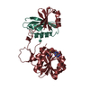
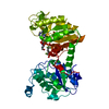
 Movie
Movie Controller


+ Open data

- Basic information
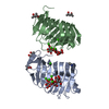
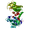
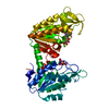
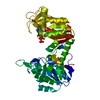
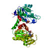
| Entry | Database: PDB / ID: 5a4r | ||||||
|---|---|---|---|---|---|---|---|
| Title | Crystal structure of a vitamin B12 trafficking protein | ||||||
 Components Components | METHYLMALONIC ACIDURIA AND HOMOCYSTINURIA TYPE D HOMOLOG, MITOCHONDRIAL | ||||||
 Keywords Keywords | TRANSPORT PROTEIN | ||||||
| Function / homology | Cobalamin (Cbl) metabolism / Methylmalonic aciduria and homocystinuria type D protein / Methylmalonic aciduria and homocystinuria type D protein / cobalamin metabolic process / mitochondrion / cytoplasm / cytosol / Cobalamin trafficking protein CblD Function and homology information Function and homology information | ||||||
| Biological species |  | ||||||
| Method |  X-RAY DIFFRACTION / SYNCHROTRON / MOLECULAR REPLACEMENT / Resolution: 2.25 Å X-RAY DIFFRACTION / SYNCHROTRON / MOLECULAR REPLACEMENT / Resolution: 2.25 Å | ||||||
 Authors Authors | Kopec, J. / Fitzpatrick, F. / Froese, D.S. / Velupillai, S. / Nowak, R. / Chalk, R. / Burgess-Brown, N. / von Delft, F. / Arrowsmith, C. / Edwards, A. ...Kopec, J. / Fitzpatrick, F. / Froese, D.S. / Velupillai, S. / Nowak, R. / Chalk, R. / Burgess-Brown, N. / von Delft, F. / Arrowsmith, C. / Edwards, A. / Bountra, C. / Fowler, B. / Baumgartner, M.R. / Yue, W.W. | ||||||
 Citation Citation | Journal: J.Biol.Chem. / Year: 2015 Title: Structural Insights Into the Mmachc-Mmadhc Protein Complex Involved in Vitamin B12 Trafficking. Authors: Froese, D.S. / Kopec, J. / Fitzpatrick, F. / Schuller, M. / Mccorvie, T.J. / Chalk, R. / Plessl, T. / Fettelschoss, V. / Fowler, B. / Baumgartner, M.R. / Yue, W.W. | ||||||
| History |
|
- Structure visualization
| Structure viewer | Molecule: MolmilJmol/JSmol |
|---|
- Downloads & links
-Download
| PDBx/mmCIF format | 5a4r.cif.gz | 41.2 KB | Display | PDBx/mmCIF format |
|---|---|---|---|---|
| PDB format | pdb5a4r.ent.gz | 28.4 KB | Display | PDB format |
| PDBx/mmJSON format | 5a4r.json.gz | Tree view | PDBx/mmJSON format | |
| Others |  Other downloads Other downloads |
-Validation report
| Arichive directory | https://data.pdbj.org/pub/pdb/validation_reports/a4/5a4rftp://data.pdbj.org/pub/pdb/validation_reports/a4/5a4r | HTTPS FTP |
|---|
-Related structure data
| Similar structure data |
|---|

-Links
 PDBj
PDBj- Assembly
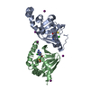
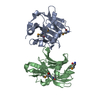
| Deposited unit | 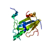
| ||||||||
|---|---|---|---|---|---|---|---|---|---|
| 1 | 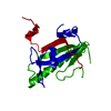
| ||||||||
| Unit cell |
|
-Components
| #1: Protein | Mass: 19215.596 Da / Num. of mol.: 1 / Fragment: RESIDUES 129-296 Source method: isolated from a genetically manipulated source Source: (gene. exp.)  |
|---|---|
| #2: Water | ChemComp-HOH /  Mass: 18.015 Da / Num. of mol.: 26 / Source method: isolated from a natural source / Formula: H2O Mass: 18.015 Da / Num. of mol.: 26 / Source method: isolated from a natural source / Formula: H2O |
-Experimental details
-Experiment
| Experiment | Method: X-RAY DIFFRACTION / Number of used crystals: 1 |
|---|
- Sample preparation
| Crystal | Density Matthews: 2.8 Å3/Da / Density % sol: 56.06 % / Description: NONE |
|---|---|
| Crystal grow | Details: 22% POLYACRYLIC ACID 5100, 0.02M MAGNESIUM CHLORIDE, 0.1M HEPES PH 7.5 |
-Data collection
| Diffraction | Mean temperature: 100 K |
|---|---|
| Diffraction source | Source: SYNCHROTRON / Site: Diamond  / Beamline: I04 / Wavelength: 0.97826 / Beamline: I04 / Wavelength: 0.97826 |
| Detector | Type: DECTRIS PILATUS / Detector: PIXEL / Date: Oct 4, 2014 |
| Radiation | Protocol: SINGLE WAVELENGTH / Monochromatic (M) / Laue (L): M / Scattering type: x-ray |
| Radiation wavelength | Wavelength: 0.97826 Å / Relative weight: 1 |
| Reflection | Resolution: 2.25→44.76 Å / Num. obs: 10530 / % possible obs: 99.8 % / Observed criterion σ(I): 1 / Redundancy: 4.9 % / Biso Wilson estimate: 55.59 Å2 / Rmerge(I) obs: 0.02 / Net I/σ(I): 31.6 |
| Reflection shell | Resolution: 2.25→2.31 Å / Redundancy: 5 % / Rmerge(I) obs: 0.6 / Mean I/σ(I) obs: 2.6 / % possible all: 99.6 |
- Processing
| Software |
| |||||||||||||||||||||||||||||||||||
|---|---|---|---|---|---|---|---|---|---|---|---|---|---|---|---|---|---|---|---|---|---|---|---|---|---|---|---|---|---|---|---|---|---|---|---|---|
| Refinement | Method to determine structure: MOLECULAR REPLACEMENT / Resolution: 2.25→44.765 Å / SU ML: 0.28 / σ(F): 1.35 / Phase error: 31.53 / Stereochemistry target values: ML Details: MISSING ELECTRON DENSITY FOR RESIDUES 129 TO 131, 160 TO 168, 237 TO 245
| |||||||||||||||||||||||||||||||||||
| Solvent computation | Shrinkage radii: 0.9 Å / VDW probe radii: 1.11 Å / Solvent model: FLAT BULK SOLVENT MODEL / Bsol: 0 Å2 / ksol: 0 e/Å3 | |||||||||||||||||||||||||||||||||||
| Displacement parameters | Biso mean: 66.2 Å2 | |||||||||||||||||||||||||||||||||||
| Refinement step | Cycle: LAST / Resolution: 2.25→44.765 Å
| |||||||||||||||||||||||||||||||||||
| Refine LS restraints |
| |||||||||||||||||||||||||||||||||||
| LS refinement shell |
|
