 Movie
Movie Controller

[English] 日本語
 Yorodumi
- PDB-4yza: C. bescii Family 3 pectate lyase double mutant K108A/Q111A in com... -
+ Open data

- Basic information
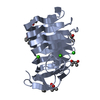
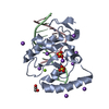
| Entry | Database: PDB / ID: 4yza | ||||||||||||
|---|---|---|---|---|---|---|---|---|---|---|---|---|---|
| Title | C. bescii Family 3 pectate lyase double mutant K108A/Q111A in complex with trigalacturonic acid | ||||||||||||
 Components Components | Pectate lyase | ||||||||||||
 Keywords Keywords | LYASE / PL3 / PARALLEL BETA-HELIX | ||||||||||||
| Function / homology |  Function and homology information Function and homology informationpectate lyase / pectate lyase activity / hydrolase activity / extracellular region / metal ion binding Similarity search - Function | ||||||||||||
| Biological species |   Caldicellulosiruptor bescii (bacteria) Caldicellulosiruptor bescii (bacteria) | ||||||||||||
| Method |  X-RAY DIFFRACTION / SYNCHROTRON / Resolution: 1.25 Å X-RAY DIFFRACTION / SYNCHROTRON / Resolution: 1.25 Å | ||||||||||||
 Authors Authors | Alahuhta, P.M. / Lunin, V.V. | ||||||||||||
| Funding support |  United States, 1items United States, 1items
| ||||||||||||
 Citation Citation | Journal: Acta Crystallogr.,Sect.D / Year: 2015 Title: The catalytic mechanism and unique low pH optimum of Caldicellulosiruptor bescii family 3 pectate lyase. Authors: Alahuhta, M. / Taylor, L.E. / Brunecky, R. / Sammond, D.W. / Michener, W. / Adams, M.W. / Himmel, M.E. / Bomble, Y.J. / Lunin, V. | ||||||||||||
| History |
|
- Structure visualization
| Structure viewer | Molecule: MolmilJmol/JSmol |
|---|
- Downloads & links
-Download
| PDBx/mmCIF format | 4yza.cif.gz | 216.8 KB | Display | PDBx/mmCIF format |
|---|---|---|---|---|
| PDB format | pdb4yza.ent.gz | Display | PDB format | |
| PDBx/mmJSON format | 4yza.json.gz | Tree view | PDBx/mmJSON format | |
| Others |  Other downloads Other downloads |
-Validation report
| Arichive directory | https://data.pdbj.org/pub/pdb/validation_reports/yz/4yzaftp://data.pdbj.org/pub/pdb/validation_reports/yz/4yza | HTTPS FTP |
|---|
-Related structure data
| Related structure data |  4yz0C  4yzqC  4yzxC  4z03C  4z05C  4z06C C: citing same article ( |
|---|---|
| Similar structure data |


-Links
 PDBj
PDBj


- Assembly
| Deposited unit | 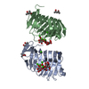
| ||||||||
|---|---|---|---|---|---|---|---|---|---|
| 1 | 
| ||||||||
| 2 | 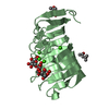
| ||||||||
| Unit cell |
| ||||||||
| Components on special symmetry positions |
|
-Components
-Protein , 1 types, 2 molecules AB
| #1: Protein | Mass: 22048.857 Da / Num. of mol.: 2 / Mutation: K375A, Q378A Source method: isolated from a genetically manipulated source Source: (gene. exp.) Caldicellulosiruptor bescii (strain ATCC BAA-1888 / DSM 6725 / Z-1320) (bacteria)Strain: ATCC BAA-1888 / DSM 6725 / Z-1320 / Gene: Athe_1854 / Production host: |
|---|
-Sugars , 4 types, 11 molecules 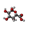

| #2: Polysaccharide | alpha-D-galactopyranuronic acid-(1-4)-alpha-D-galactopyranuronic acid-(1-4)-alpha-D-talopyranuronic acid Source method: isolated from a genetically manipulated source | ||
|---|---|---|---|
| #3: Polysaccharide | alpha-D-galactopyranuronic acid-(1-4)-alpha-D-galactopyranuronic acid-(1-4)-alpha-D-galactopyranuronic acid Source method: isolated from a genetically manipulated source | ||
| #8: Sugar | ChemComp-ADA /  Type: D-saccharide, alpha linking / Mass: 194.139 Da / Num. of mol.: 7 Type: D-saccharide, alpha linking / Mass: 194.139 Da / Num. of mol.: 7Source method: isolated from a genetically manipulated source Formula: C6H10O7 #9: Sugar |  Type: D-saccharide, beta linking / Mass: 194.139 Da / Num. of mol.: 2 Type: D-saccharide, beta linking / Mass: 194.139 Da / Num. of mol.: 2Source method: isolated from a genetically manipulated source Formula: C6H10O7 |
-Non-polymers , 5 types, 626 molecules 


| #4: Chemical | ChemComp-CA /  Mass: 40.078 Da / Num. of mol.: 9 / Source method: obtained synthetically / Formula: Ca Mass: 40.078 Da / Num. of mol.: 9 / Source method: obtained synthetically / Formula: Ca#5: Chemical | ChemComp-MPD / (  Mass: 118.174 Da / Num. of mol.: 4 / Source method: obtained synthetically / Formula: C6H14O2 / Comment: precipitant*YM Mass: 118.174 Da / Num. of mol.: 4 / Source method: obtained synthetically / Formula: C6H14O2 / Comment: precipitant*YM#6: Chemical | ChemComp-MRD / ( |  Mass: 118.174 Da / Num. of mol.: 1 / Source method: obtained synthetically / Formula: C6H14O2 / Comment: precipitant*YM Mass: 118.174 Da / Num. of mol.: 1 / Source method: obtained synthetically / Formula: C6H14O2 / Comment: precipitant*YM#7: Chemical | ChemComp-IMD / |  Mass: 69.085 Da / Num. of mol.: 1 / Source method: obtained synthetically / Formula: C3H5N2 Mass: 69.085 Da / Num. of mol.: 1 / Source method: obtained synthetically / Formula: C3H5N2#10: Water | ChemComp-HOH / | Mass: 18.015 Da / Num. of mol.: 611 / Source method: isolated from a natural source / Formula: H2O |
|---|
-Details
| Has protein modification | N |
|---|
-Experimental details
-Experiment
| Experiment | Method: X-RAY DIFFRACTION |
|---|
- Sample preparation
| Crystal | Density Matthews: 2.11 Å3/Da / Density % sol: 41.79 % |
|---|---|
| Crystal grow | Temperature: 293 K / Method: vapor diffusion, sitting drop / Details: 0.1 M Tris pH 9 and 65.9% MPD |
-Data collection
| Diffraction | Mean temperature: 100 K |
|---|---|
| Diffraction source | Source: SYNCHROTRON / Site: SSRL / Beamline: BL11-1 / Wavelength: 0.98397 Å |
| Detector | Type: Bruker Platinum 135 / Detector: CCD / Date: Mar 4, 2014 |
| Radiation | Protocol: SINGLE WAVELENGTH / Monochromatic (M) / Laue (L): M / Scattering type: x-ray |
| Radiation wavelength | Wavelength: 0.98397 Å / Relative weight: 1 |
| Reflection | Resolution: 1.25→50.89 Å / Num. obs: 100777 / % possible obs: 98.5 % / Redundancy: 11.6 % / Rmerge(I) obs: 0.052 / Net I/σ(I): 25.6 |
| Reflection shell | Resolution: 1.25→1.27 Å / Redundancy: 6.1 % / Rmerge(I) obs: 0.782 / Mean I/σ(I) obs: 2.3 / % possible all: 95 |
- Processing
| Software |
| ||||||||||||||||||||||||||||||||||||||||||||||||||||||||||||||||||||||||||||||||||||||||||||||||||||||||||||||||||||||||||||||||||||||||||||||||||||||||||||||||||||||||||||||||||||||
|---|---|---|---|---|---|---|---|---|---|---|---|---|---|---|---|---|---|---|---|---|---|---|---|---|---|---|---|---|---|---|---|---|---|---|---|---|---|---|---|---|---|---|---|---|---|---|---|---|---|---|---|---|---|---|---|---|---|---|---|---|---|---|---|---|---|---|---|---|---|---|---|---|---|---|---|---|---|---|---|---|---|---|---|---|---|---|---|---|---|---|---|---|---|---|---|---|---|---|---|---|---|---|---|---|---|---|---|---|---|---|---|---|---|---|---|---|---|---|---|---|---|---|---|---|---|---|---|---|---|---|---|---|---|---|---|---|---|---|---|---|---|---|---|---|---|---|---|---|---|---|---|---|---|---|---|---|---|---|---|---|---|---|---|---|---|---|---|---|---|---|---|---|---|---|---|---|---|---|---|---|---|---|---|
| Refinement | Resolution: 1.25→50.89 Å / Cor.coef. Fo:Fc: 0.987 / Cor.coef. Fo:Fc free: 0.974 / SU B: 1.523 / SU ML: 0.029 / Cross valid method: THROUGHOUT / ESU R: 0.037 / ESU R Free: 0.041 / Stereochemistry target values: MAXIMUM LIKELIHOOD / Details: HYDROGENS HAVE BEEN ADDED IN THE RIDING POSITIONS
| ||||||||||||||||||||||||||||||||||||||||||||||||||||||||||||||||||||||||||||||||||||||||||||||||||||||||||||||||||||||||||||||||||||||||||||||||||||||||||||||||||||||||||||||||||||||
| Solvent computation | Ion probe radii: 0.8 Å / Shrinkage radii: 0.8 Å / VDW probe radii: 1.2 Å / Solvent model: MASK | ||||||||||||||||||||||||||||||||||||||||||||||||||||||||||||||||||||||||||||||||||||||||||||||||||||||||||||||||||||||||||||||||||||||||||||||||||||||||||||||||||||||||||||||||||||||
| Displacement parameters | Biso mean: 15.14 Å2
| ||||||||||||||||||||||||||||||||||||||||||||||||||||||||||||||||||||||||||||||||||||||||||||||||||||||||||||||||||||||||||||||||||||||||||||||||||||||||||||||||||||||||||||||||||||||
| Refinement step | Cycle: LAST / Resolution: 1.25→50.89 Å
| ||||||||||||||||||||||||||||||||||||||||||||||||||||||||||||||||||||||||||||||||||||||||||||||||||||||||||||||||||||||||||||||||||||||||||||||||||||||||||||||||||||||||||||||||||||||
| Refine LS restraints |
|