Method to determine structure: SIRAS Starting model: NONE Resolution: 1.699→34.215 Å / SU ML: 0.17 / σ(F): 1.36 / Phase error: 19.38 / Stereochemistry target values: ML Details: THE OCCUPANCY OF SIDE CHAIN ATOMS, FOR WHICH ELECTRON DENSITY WAS POOR, IS SET TO 0.
Rfactor
Num. reflection
% reflection
Rfree
0.1945
2384
7.8 %
Rwork
0.1663
-
-
obs
0.1685
30656
99.6 %
Solvent computation
Shrinkage radii: 0.9 Å / VDW probe radii: 1.11 Å / Solvent model: FLAT BULK SOLVENT MODEL
Refinement step
Cycle: LAST / Resolution: 1.699→34.215 Å
Protein
Nucleic acid
Ligand
Solvent
Total
Num. atoms
1678
0
30
258
1966
Refine LS restraints
Refine-ID
Type
Dev ideal
Number
X-RAY DIFFRACTION
f_bond_d
0.007
1759
X-RAY DIFFRACTION
f_angle_d
1.046
2375
X-RAY DIFFRACTION
f_dihedral_angle_d
13.118
666
X-RAY DIFFRACTION
f_chiral_restr
0.077
260
X-RAY DIFFRACTION
f_plane_restr
0.004
301
LS refinement shell
Resolution (Å)
Rfactor Rfree
Num. reflection Rfree
Rfactor Rwork
Num. reflection Rwork
Refine-ID
% reflection obs (%)
1.6991-1.7338
0.2663
131
0.2948
1600
X-RAY DIFFRACTION
97
1.7338-1.7715
0.3417
142
0.2732
1618
X-RAY DIFFRACTION
99
1.7715-1.8127
0.2982
136
0.2519
1635
X-RAY DIFFRACTION
99
1.8127-1.858
0.2909
139
0.2289
1634
X-RAY DIFFRACTION
99
1.858-1.9083
0.2363
136
0.2126
1661
X-RAY DIFFRACTION
99
1.9083-1.9644
0.2379
139
0.1966
1614
X-RAY DIFFRACTION
100
1.9644-2.0278
0.2066
142
0.1798
1652
X-RAY DIFFRACTION
100
2.0278-2.1003
0.2102
138
0.1647
1656
X-RAY DIFFRACTION
100
2.1003-2.1844
0.1965
138
0.1533
1660
X-RAY DIFFRACTION
100
2.1844-2.2837
0.1717
140
0.1535
1662
X-RAY DIFFRACTION
100
2.2837-2.4041
0.1732
137
0.1454
1644
X-RAY DIFFRACTION
100
2.4041-2.5547
0.1895
146
0.1513
1675
X-RAY DIFFRACTION
100
2.5547-2.7519
0.1846
140
0.1562
1670
X-RAY DIFFRACTION
100
2.7519-3.0287
0.1946
141
0.161
1678
X-RAY DIFFRACTION
100
3.0287-3.4665
0.174
140
0.1475
1711
X-RAY DIFFRACTION
100
3.4665-4.3661
0.1474
143
0.1331
1709
X-RAY DIFFRACTION
100
4.3661-34.2219
0.1943
156
0.1739
1793
X-RAY DIFFRACTION
100
Refinement TLS params.
Method: refined / Refine-ID: X-RAY DIFFRACTION
ID
L11 (°2)
L12 (°2)
L13 (°2)
L22 (°2)
L23 (°2)
L33 (°2)
S11 (Å °)
S12 (Å °)
S13 (Å °)
S21 (Å °)
S22 (Å °)
S23 (Å °)
S31 (Å °)
S32 (Å °)
S33 (Å °)
T11 (Å2)
T12 (Å2)
T13 (Å2)
T22 (Å2)
T23 (Å2)
T33 (Å2)
Origin x (Å)
Origin y (Å)
Origin z (Å)
1
1.0979
0.1448
0.1102
1.1729
-0.2884
1.5833
0.0296
0.0371
0.0126
0.0015
0.02
0.0954
0.0467
-0.2957
-0.0633
0.112
0.0054
-0.002
0.134
0.0065
0.0928
28.084
19.6978
39.1085
2
1.3279
0.3918
-0.0641
0.8878
0.1502
2.0502
0.0212
0.0208
0.1607
-0.0124
0.0315
0.0476
-0.2796
0.022
-0.042
0.1191
0.0014
-0.0024
0.0549
-0.0031
0.0884
38.7594
27.5011
49.7764
3
0.5194
0.1139
-0.2727
0.3234
0.1819
1.6968
-0.0039
-0.0426
-0.0831
0.0588
0.0421
-0.1018
0.2338
0.254
-0.0541
0.1295
0.038
-0.0225
0.1277
-0.013
0.1322
44.6407
13.8188
46.7265
Refinement TLS group
ID
Refine-ID
Refine TLS-ID
Selection details
1
X-RAY DIFFRACTION
1
CHAIN 'A' AND (RESID399THROUGH460 )
2
X-RAY DIFFRACTION
2
CHAIN 'A' AND (RESID461THROUGH546 )
3
X-RAY DIFFRACTION
3
CHAIN 'A' AND (RESID547THROUGH606 )
+
About Yorodumi
-
News
-
Feb 9, 2022. New format data for meta-information of EMDB entries
New format data for meta-information of EMDB entries
Version 3 of the EMDB header file is now the official format.
The previous official version 1.9 will be removed from the archive.
In the structure databanks used in Yorodumi, some data are registered as the other names, "COVID-19 virus" and "2019-nCoV". Here are the details of the virus and the list of structure data.
Jan 31, 2019. EMDB accession codes are about to change! (news from PDBe EMDB page)
EMDB accession codes are about to change! (news from PDBe EMDB page)
The allocation of 4 digits for EMDB accession codes will soon come to an end. Whilst these codes will remain in use, new EMDB accession codes will include an additional digit and will expand incrementally as the available range of codes is exhausted. The current 4-digit format prefixed with “EMD-” (i.e. EMD-XXXX) will advance to a 5-digit format (i.e. EMD-XXXXX), and so on. It is currently estimated that the 4-digit codes will be depleted around Spring 2019, at which point the 5-digit format will come into force.
The EM Navigator/Yorodumi systems omit the EMD- prefix.
Related info.:Q: What is EMD? / ID/Accession-code notation in Yorodumi/EM Navigator
Yorodumi is a browser for structure data from EMDB, PDB, SASBDB, etc.
This page is also the successor to EM Navigator detail page, and also detail information page/front-end page for Omokage search.
The word "yorodu" (or yorozu) is an old Japanese word meaning "ten thousand". "mi" (miru) is to see.
Related info.:EMDB / PDB / SASBDB / Comparison of 3 databanks / Yorodumi Search / Aug 31, 2016. New EM Navigator & Yorodumi / Yorodumi Papers / Jmol/JSmol / Function and homology information / Changes in new EM Navigator and Yorodumi
 Movie
Movie Controller


 Open data
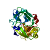
 Basic information
 Components
 Keywords
 Function and homology information
 X-RAY DIFFRACTION /
  Authors
 Citation
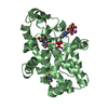
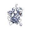
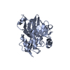
 Structure visualization
 Downloads & links
 Other downloads

 PDBj
PDBj Assembly


 Mass: 96.063 Da / Num. of mol.: 6 / Source method: obtained synthetically / Formula: SO4
 Mass: 18.015 Da / Num. of mol.: 258 / Source method: isolated from a natural source / Formula: H2O
 Sample preparation
 / Beamline: X06DA / Wavelength: 0.99986
  Processing