 Movie
Movie Controller

[English] 日本語
 Yorodumi
- PDB-5a0v: Catalysis and 5' end sensing by ribonuclease RNase J of the metal... -
+ Open data

- Basic information
| Entry | Database: PDB / ID: 5a0v | ||||||
|---|---|---|---|---|---|---|---|
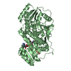
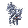
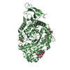
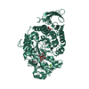
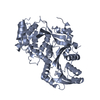
| Title | Catalysis and 5' end sensing by ribonuclease RNase J of the metallo- beta-lactamase family | ||||||
 Components Components |
| ||||||
 Keywords Keywords | HYDROLASE/RNA / HYDROLASE-RNA COMPLEX / ENDONUCLEASE / EXONUCLEASE | ||||||
| Function / homology |  Function and homology information Function and homology informationRNA exonuclease activity / 5'-3' RNA exonuclease activity / 5'-3' exonuclease activity / RNA endonuclease activity / mRNA processing / rRNA processing / Hydrolases; Acting on ester bonds / RNA binding / zinc ion binding / cytoplasm Similarity search - Function | ||||||
| Biological species | STREPTOMYCES COELICOLOR A3 | ||||||
| Method |  X-RAY DIFFRACTION / SYNCHROTRON / MOLECULAR REPLACEMENT / Resolution: 2.8 Å X-RAY DIFFRACTION / SYNCHROTRON / MOLECULAR REPLACEMENT / Resolution: 2.8 Å | ||||||
 Authors Authors | Pei, X.Y. / Bralley, P. / Jones, G.H. / Luisi, B.F. | ||||||
 Citation Citation | Journal: Nucleic Acids Res. / Year: 2015 Title: Linkage of Catalysis and 5' End Recognition in Ribonuclease Rnase J Authors: Pei, X.Y. / Bralley, P. / Jones, G.H. / Luisi, B.F. | ||||||
| History |
|
- Structure visualization
| Structure viewer | Molecule: MolmilJmol/JSmol |
|---|
- Downloads & links
-Download
| PDBx/mmCIF format | 5a0v.cif.gz | 385.2 KB | Display | PDBx/mmCIF format |
|---|---|---|---|---|
| PDB format | pdb5a0v.ent.gz | 314.1 KB | Display | PDB format |
| PDBx/mmJSON format | 5a0v.json.gz | Tree view | PDBx/mmJSON format | |
| Others |  Other downloads Other downloads |
-Validation report
| Arichive directory | https://data.pdbj.org/pub/pdb/validation_reports/a0/5a0vftp://data.pdbj.org/pub/pdb/validation_reports/a0/5a0v | HTTPS FTP |
|---|
-Related structure data
| Related structure data | 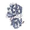 5a0tSC S: Starting model for refinement C: citing same article ( |
|---|---|
| Similar structure data |

-Links
 PDBj
PDBj

- Assembly
| Deposited unit | 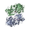
| ||||||||
|---|---|---|---|---|---|---|---|---|---|
| 1 | 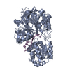
| ||||||||
| 2 | 
| ||||||||
| Unit cell |
| ||||||||
| Noncrystallographic symmetry (NCS) | NCS oper: (Code: given Matrix: (0.2866, -0.000552, -0.9581), Vector: |
-Components
| #1: Protein | Mass: 61146.090 Da / Num. of mol.: 2 Fragment: BETA-LACTAMASE DOMAIN AND BETA-CASP DOMAIN, RESIDUES 1-561 Source method: isolated from a genetically manipulated source Source: (gene. exp.)  STREPTOMYCES COELICOLOR A3(2) (bacteria) STREPTOMYCES COELICOLOR A3(2) (bacteria)Plasmid: PET19B / Production host: #2: RNA chain | Mass: 1827.141 Da / Num. of mol.: 2 Source method: isolated from a genetically manipulated source Source: (gene. exp.) STREPTOMYCES COELICOLOR A3(2) (bacteria)Plasmid: PET19B / Production host: #3: Chemical | ChemComp-ZN /   Mass: 65.409 Da / Num. of mol.: 4 / Source method: obtained synthetically / Formula: Zn Mass: 65.409 Da / Num. of mol.: 4 / Source method: obtained synthetically / Formula: Zn#4: Chemical | ChemComp-C5P / |   Mass: 323.197 Da / Num. of mol.: 1 / Source method: obtained synthetically / Formula: C9H14N3O8P Mass: 323.197 Da / Num. of mol.: 1 / Source method: obtained synthetically / Formula: C9H14N3O8P#5: Water | ChemComp-HOH / |  Mass: 18.015 Da / Num. of mol.: 75 / Source method: isolated from a natural source / Formula: H2O Mass: 18.015 Da / Num. of mol.: 75 / Source method: isolated from a natural source / Formula: H2O |
|---|
-Experimental details
-Experiment
| Experiment | Method: X-RAY DIFFRACTION / Number of used crystals: 1 |
|---|
- Sample preparation
| Crystal | Density Matthews: 4.9 Å3/Da / Density % sol: 0.74 % Description: DATA WERE COLLECTED USING THE OSCILLATION METHOD |
|---|---|
| Crystal grow | pH: 8.5 Details: THE BEST CRYSTALS OF S. COELICOLOR RNASE J WERE OBTAINED IN THE 27.5% W/V PEG 400, 0.1 M TRIS-HCL, PH 8.5 BY MIXING A 1:2 VOLUME RATIO OF CRYSTALLIZATION RESERVOIR TO PROTEIN SOLUTION, THEN SOAKED WITH MNCL2 |
-Data collection
| Diffraction | Mean temperature: 100 K |
|---|---|
| Diffraction source | Source: SYNCHROTRON / Site: Diamond  / Beamline: I04-1 / Wavelength: 0.91742 / Beamline: I04-1 / Wavelength: 0.91742 |
| Detector | Type: DECTRIS PILATUS / Detector: PIXEL / Date: Feb 1, 2015 |
| Radiation | Protocol: SINGLE WAVELENGTH / Monochromatic (M) / Laue (L): M / Scattering type: x-ray |
| Radiation wavelength | Wavelength: 0.91742 Å / Relative weight: 1 |
| Reflection | Resolution: 2.7→2.75 Å / Num. obs: 47608 / % possible obs: 99.8 % / Observed criterion σ(I): 2.5 / Redundancy: 6.9 % / Biso Wilson estimate: 55.49 Å2 / Rmerge(I) obs: 0.14 / Net I/σ(I): 9.7 |
| Reflection shell | Resolution: 2.7→2.85 Å / Redundancy: 14.4 % / Rmerge(I) obs: 0.77 / Mean I/σ(I) obs: 2.5 / % possible all: 99.5 |
- Processing
| Software |
| |||||||||||||||||||||||||||||||||||||||||||||||||||||||||||||||||||||||||||||||||||||||||||||||||||||||||||||||||||||||||||||||||||||||||||||||||||||||||||||||||||||||||||||||||||||||||||||||||||||||||||||||||||||||||
|---|---|---|---|---|---|---|---|---|---|---|---|---|---|---|---|---|---|---|---|---|---|---|---|---|---|---|---|---|---|---|---|---|---|---|---|---|---|---|---|---|---|---|---|---|---|---|---|---|---|---|---|---|---|---|---|---|---|---|---|---|---|---|---|---|---|---|---|---|---|---|---|---|---|---|---|---|---|---|---|---|---|---|---|---|---|---|---|---|---|---|---|---|---|---|---|---|---|---|---|---|---|---|---|---|---|---|---|---|---|---|---|---|---|---|---|---|---|---|---|---|---|---|---|---|---|---|---|---|---|---|---|---|---|---|---|---|---|---|---|---|---|---|---|---|---|---|---|---|---|---|---|---|---|---|---|---|---|---|---|---|---|---|---|---|---|---|---|---|---|---|---|---|---|---|---|---|---|---|---|---|---|---|---|---|---|---|---|---|---|---|---|---|---|---|---|---|---|---|---|---|---|---|---|---|---|---|---|---|---|---|---|---|---|---|---|---|---|---|
| Refinement | Method to determine structure: MOLECULAR REPLACEMENT Starting model: PDB ENTRY 5A0T Resolution: 2.8→32.719 Å / SU ML: 0.32 / σ(F): 0.05 / Phase error: 20.92 / Stereochemistry target values: ML Details: DISORDERED REGIONS OF RESIDUES 456 TO 561 IN BOTH A CHAIN AND B CHAIN WERE NOT MODELLED.
| |||||||||||||||||||||||||||||||||||||||||||||||||||||||||||||||||||||||||||||||||||||||||||||||||||||||||||||||||||||||||||||||||||||||||||||||||||||||||||||||||||||||||||||||||||||||||||||||||||||||||||||||||||||||||
| Solvent computation | Shrinkage radii: 0.9 Å / VDW probe radii: 1.11 Å / Solvent model: FLAT BULK SOLVENT MODEL | |||||||||||||||||||||||||||||||||||||||||||||||||||||||||||||||||||||||||||||||||||||||||||||||||||||||||||||||||||||||||||||||||||||||||||||||||||||||||||||||||||||||||||||||||||||||||||||||||||||||||||||||||||||||||
| Refinement step | Cycle: LAST / Resolution: 2.8→32.719 Å
| |||||||||||||||||||||||||||||||||||||||||||||||||||||||||||||||||||||||||||||||||||||||||||||||||||||||||||||||||||||||||||||||||||||||||||||||||||||||||||||||||||||||||||||||||||||||||||||||||||||||||||||||||||||||||
| Refine LS restraints |
| |||||||||||||||||||||||||||||||||||||||||||||||||||||||||||||||||||||||||||||||||||||||||||||||||||||||||||||||||||||||||||||||||||||||||||||||||||||||||||||||||||||||||||||||||||||||||||||||||||||||||||||||||||||||||
| LS refinement shell |
| |||||||||||||||||||||||||||||||||||||||||||||||||||||||||||||||||||||||||||||||||||||||||||||||||||||||||||||||||||||||||||||||||||||||||||||||||||||||||||||||||||||||||||||||||||||||||||||||||||||||||||||||||||||||||
| Refinement TLS params. | Method: refined / Origin x: 16.8119 Å / Origin y: 60.5149 Å / Origin z: -12.4589 Å
| |||||||||||||||||||||||||||||||||||||||||||||||||||||||||||||||||||||||||||||||||||||||||||||||||||||||||||||||||||||||||||||||||||||||||||||||||||||||||||||||||||||||||||||||||||||||||||||||||||||||||||||||||||||||||
| Refinement TLS group | Selection details: ALL |
