 Movie
Movie Controller


+ Open data

- Basic information
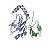
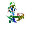
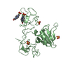
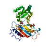
| Entry | Database: PDB / ID: 5a0o | ||||||
|---|---|---|---|---|---|---|---|
| Title | adhiron raised against p300 | ||||||
 Components Components | ADHIRON | ||||||
 Keywords Keywords | DE NOVO PROTEIN / AFFIMER | ||||||
| Function / homology | Nuclear Transport Factor 2; Chain: A, - #10 / Nuclear Transport Factor 2; Chain: A, / Roll / Alpha Beta Function and homology information Function and homology information | ||||||
| Biological species | SYNTHETIC CONSTRUCT (others) | ||||||
| Method |  X-RAY DIFFRACTION / SYNCHROTRON / MOLECULAR REPLACEMENT / Resolution: 2.73 Å X-RAY DIFFRACTION / SYNCHROTRON / MOLECULAR REPLACEMENT / Resolution: 2.73 Å | ||||||
 Authors Authors | Kyle, H.F. / Wickson, K.F. / Stott, J. / Burslem, G.M. / Breeze, A.L. / Tiede, C. / Tomlinson, D.C. / Warriner, S.L. / Nelson, A. / Wilson, A.J. / Edwards, T.A. | ||||||
 Citation Citation | Journal: Mol.Biosyst. / Year: 2015 Title: Exploration of the Hif-1Alpha.P300 Interface Using Peptide and Adhiron Phage Display Technologies Authors: Kyle, H.F. / Wickson, K.F. / Stott, J. / Burslem, G.M. / Breeze, A.L. / Tiede, C. / Tomlinson, D.C. / Warriner, S.L. / Nelson, A. / Wilson, A.J. / Edwards, T.A. | ||||||
| History |
|
- Structure visualization
| Structure viewer | Molecule: MolmilJmol/JSmol |
|---|
- Downloads & links
-Download
| PDBx/mmCIF format | 5a0o.cif.gz | 83.4 KB | Display | PDBx/mmCIF format |
|---|---|---|---|---|
| PDB format | pdb5a0o.ent.gz | 66 KB | Display | PDB format |
| PDBx/mmJSON format | 5a0o.json.gz | Tree view | PDBx/mmJSON format | |
| Others |  Other downloads Other downloads |
-Validation report
| Arichive directory | https://data.pdbj.org/pub/pdb/validation_reports/a0/5a0oftp://data.pdbj.org/pub/pdb/validation_reports/a0/5a0o | HTTPS FTP |
|---|
-Related structure data
| Related structure data |  4n6uS S: Starting model for refinement |
|---|---|
| Similar structure data |


-Links
 PDBj
PDBj
- Assembly
| Deposited unit | 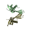
| ||||||||||||
|---|---|---|---|---|---|---|---|---|---|---|---|---|---|
| 1 |
| ||||||||||||
| Unit cell |
| ||||||||||||
| Components on special symmetry positions |
|
-Components
| #1: Protein | Mass: 12465.246 Da / Num. of mol.: 2 / Fragment: ADHIRON Source method: isolated from a genetically manipulated source Source: (gene. exp.) SYNTHETIC CONSTRUCT (others) / Description: DESIGNED NON-ANTIBODY BINDING PROTEIN / Plasmid: PET11 / Production host:  #2: Water | ChemComp-HOH / |  Mass: 18.015 Da / Num. of mol.: 48 / Source method: isolated from a natural source / Formula: H2O Mass: 18.015 Da / Num. of mol.: 48 / Source method: isolated from a natural source / Formula: H2O |
|---|
-Experimental details
-Experiment
| Experiment | Method: X-RAY DIFFRACTION / Number of used crystals: 1 |
|---|
- Sample preparation
| Crystal | Density Matthews: 3.67 Å3/Da / Density % sol: 66.5 % / Description: NONE |
|---|---|
| Crystal grow | pH: 7 / Details: 0.8 M DI-SODIUM SUCCINATE PH7 |
-Data collection
| Diffraction | Mean temperature: 100 K |
|---|---|
| Diffraction source | Source: SYNCHROTRON / Site: Diamond  / Beamline: I03 / Wavelength: 0.91741 / Beamline: I03 / Wavelength: 0.91741 |
| Detector | Type: DECTRIS PILATUS / Detector: PIXEL / Date: Feb 4, 2015 |
| Radiation | Protocol: SINGLE WAVELENGTH / Monochromatic (M) / Laue (L): M / Scattering type: x-ray |
| Radiation wavelength | Wavelength: 0.91741 Å / Relative weight: 1 |
| Reflection | Resolution: 2.73→36.73 Å / Num. obs: 7134 / % possible obs: 99.8 % / Observed criterion σ(I): 0 / Redundancy: 12.6 % / Rmerge(I) obs: 0.15 / Net I/σ(I): 12.3 |
| Reflection shell | Resolution: 2.73→2.8 Å / Redundancy: 13.8 % / Rmerge(I) obs: 1.44 / Mean I/σ(I) obs: 1.7 / % possible all: 100 |
- Processing
| Software |
| ||||||||||||||||||||||||||||||||||||||||||||||||||||||||||||||||||||||||||||||||||||||||||||||||||||||||||||||||||||||||||||||||||||||||||||||||||||||||||||||||||||||||||||||||||||||
|---|---|---|---|---|---|---|---|---|---|---|---|---|---|---|---|---|---|---|---|---|---|---|---|---|---|---|---|---|---|---|---|---|---|---|---|---|---|---|---|---|---|---|---|---|---|---|---|---|---|---|---|---|---|---|---|---|---|---|---|---|---|---|---|---|---|---|---|---|---|---|---|---|---|---|---|---|---|---|---|---|---|---|---|---|---|---|---|---|---|---|---|---|---|---|---|---|---|---|---|---|---|---|---|---|---|---|---|---|---|---|---|---|---|---|---|---|---|---|---|---|---|---|---|---|---|---|---|---|---|---|---|---|---|---|---|---|---|---|---|---|---|---|---|---|---|---|---|---|---|---|---|---|---|---|---|---|---|---|---|---|---|---|---|---|---|---|---|---|---|---|---|---|---|---|---|---|---|---|---|---|---|---|---|
| Refinement | Method to determine structure: MOLECULAR REPLACEMENT Starting model: PDB ENTRY 4N6U Resolution: 2.73→36.73 Å / Cor.coef. Fo:Fc: 0.931 / Cor.coef. Fo:Fc free: 0.911 / SU B: 14.875 / SU ML: 0.294 / Cross valid method: THROUGHOUT / ESU R: 0.91 / ESU R Free: 0.363 / Stereochemistry target values: MAXIMUM LIKELIHOOD Details: HYDROGENS HAVE BEEN ADDED IN THE RIDING POSITIONS. U VALUES REFINED INDIVIDUALLY
| ||||||||||||||||||||||||||||||||||||||||||||||||||||||||||||||||||||||||||||||||||||||||||||||||||||||||||||||||||||||||||||||||||||||||||||||||||||||||||||||||||||||||||||||||||||||
| Solvent computation | Ion probe radii: 0.8 Å / Shrinkage radii: 0.8 Å / VDW probe radii: 1.2 Å / Solvent model: MASK | ||||||||||||||||||||||||||||||||||||||||||||||||||||||||||||||||||||||||||||||||||||||||||||||||||||||||||||||||||||||||||||||||||||||||||||||||||||||||||||||||||||||||||||||||||||||
| Displacement parameters | Biso mean: 70.371 Å2
| ||||||||||||||||||||||||||||||||||||||||||||||||||||||||||||||||||||||||||||||||||||||||||||||||||||||||||||||||||||||||||||||||||||||||||||||||||||||||||||||||||||||||||||||||||||||
| Refinement step | Cycle: LAST / Resolution: 2.73→36.73 Å
| ||||||||||||||||||||||||||||||||||||||||||||||||||||||||||||||||||||||||||||||||||||||||||||||||||||||||||||||||||||||||||||||||||||||||||||||||||||||||||||||||||||||||||||||||||||||
| Refine LS restraints |
|