 Movie
Movie Controller

[English] 日本語
 Yorodumi
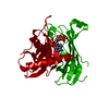
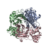
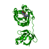
- PDB-4zv3: Crystal structure of the N- and C-terminal domains of mouse acyl-... -
+ Open data

- Basic information
| Entry | Database: PDB / ID: 4zv3 | ||||||
|---|---|---|---|---|---|---|---|
| Title | Crystal structure of the N- and C-terminal domains of mouse acyl-CoA thioesterase 7 | ||||||
 Components Components | Cytosolic acyl coenzyme A thioester hydrolase | ||||||
 Keywords Keywords | HYDROLASE / Thioesterase / Double hotdog / Inflammation | ||||||
| Function / homology |  Function and homology information Function and homology informationlong-chain fatty-acyl-CoA catabolic process / medium-chain fatty acid biosynthetic process / long-chain fatty acyl-CoA binding / palmitic acid biosynthetic process / palmitoyl-CoA hydrolase / medium-chain fatty-acyl-CoA catabolic process / long-chain fatty acyl-CoA hydrolase activity / acyl-CoA metabolic process / fatty-acyl-CoA binding / fatty acyl-CoA hydrolase activity ...long-chain fatty-acyl-CoA catabolic process / medium-chain fatty acid biosynthetic process / long-chain fatty acyl-CoA binding / palmitic acid biosynthetic process / palmitoyl-CoA hydrolase / medium-chain fatty-acyl-CoA catabolic process / long-chain fatty acyl-CoA hydrolase activity / acyl-CoA metabolic process / fatty-acyl-CoA binding / fatty acyl-CoA hydrolase activity / Mitochondrial Fatty Acid Beta-Oxidation / fatty acid catabolic process / coenzyme A biosynthetic process / carboxylic ester hydrolase activity / cell body / neuron projection / mitochondrial matrix / protein homodimerization activity / nucleoplasm / identical protein binding / cytoplasm / cytosol Similarity search - Function | ||||||
| Biological species |  | ||||||
| Method |  X-RAY DIFFRACTION / SYNCHROTRON / MOLECULAR REPLACEMENT / Resolution: 3.1 Å X-RAY DIFFRACTION / SYNCHROTRON / MOLECULAR REPLACEMENT / Resolution: 3.1 Å | ||||||
 Authors Authors | Swarbrick, C.M.D. / Forwood, J.K. | ||||||
 Citation Citation | Journal: To Be Published Title: Crystal structure of the N- and C-terminal domains of mouse acyl-CoA thioesterase 7 Authors: Swarbrick, C.M.D. / Forwood, J.K. | ||||||
| History |
|
- Structure visualization
| Structure viewer | Molecule: MolmilJmol/JSmol |
|---|
- Downloads & links
-Download
| PDBx/mmCIF format | 4zv3.cif.gz | 188 KB | Display | PDBx/mmCIF format |
|---|---|---|---|---|
| PDB format | pdb4zv3.ent.gz | 150.3 KB | Display | PDB format |
| PDBx/mmJSON format | 4zv3.json.gz | Tree view | PDBx/mmJSON format | |
| Others |  Other downloads Other downloads |
-Validation report
| Arichive directory | https://data.pdbj.org/pub/pdb/validation_reports/zv/4zv3ftp://data.pdbj.org/pub/pdb/validation_reports/zv/4zv3 | HTTPS FTP |
|---|
-Related structure data


-Links
 PDBj
PDBj


- Assembly
| Deposited unit | 
| ||||||||
|---|---|---|---|---|---|---|---|---|---|
| 1 |
| ||||||||
| Unit cell |
|
-Components
| #1: Protein | Mass: 35417.508 Da / Num. of mol.: 3 / Fragment: unp residues 55-369 Source method: isolated from a genetically manipulated source Source: (gene. exp.)  #2: Chemical |   Mass: 767.534 Da / Num. of mol.: 3 / Source method: obtained synthetically / Formula: C21H36N7O16P3S Mass: 767.534 Da / Num. of mol.: 3 / Source method: obtained synthetically / Formula: C21H36N7O16P3S |
|---|
-Experimental details
-Experiment
| Experiment | Method: X-RAY DIFFRACTION |
|---|
- Sample preparation
| Crystal | Density Matthews: 2.58 Å3/Da / Density % sol: 52.39 % |
|---|---|
| Crystal grow | Temperature: 296 K / Method: vapor diffusion, hanging drop / Details: 200 mM sodium citrate, 12% PEG 3350 |
-Data collection
| Diffraction | Mean temperature: 100 K |
|---|---|
| Diffraction source | Source: SYNCHROTRON / Site: Australian Synchrotron  / Beamline: MX2 / Wavelength: 0.9537 Å / Beamline: MX2 / Wavelength: 0.9537 Å |
| Detector | Type: ADSC QUANTUM 315r / Detector: CCD / Date: Aug 13, 2014 |
| Radiation | Protocol: SINGLE WAVELENGTH / Monochromatic (M) / Laue (L): M / Scattering type: x-ray |
| Radiation wavelength | Wavelength: 0.9537 Å / Relative weight: 1 |
| Reflection | Resolution: 3.1→34.91 Å / Num. obs: 18941 / % possible obs: 99.9 % / Redundancy: 3.3 % / Rmerge(I) obs: 0.157 / Net I/σ(I): 5.5 |
- Processing
| Software |
| ||||||||||||||||||||
|---|---|---|---|---|---|---|---|---|---|---|---|---|---|---|---|---|---|---|---|---|---|
| Refinement | Method to determine structure: MOLECULAR REPLACEMENT Starting model: 2Q2B, 2V1O Resolution: 3.1→32.1 Å / Cor.coef. Fo:Fc: 0.911 / Cor.coef. Fo:Fc free: 0.872 / SU B: 0.015 / SU ML: 0 / Cross valid method: THROUGHOUT / ESU R: 0.479 / ESU R Free: 0.589 / Stereochemistry target values: MAXIMUM LIKELIHOOD / Details: HYDROGENS HAVE BEEN ADDED IN THE RIDING POSITIONS
| ||||||||||||||||||||
| Solvent computation | Ion probe radii: 0.8 Å / Shrinkage radii: 0.8 Å / VDW probe radii: 1.2 Å / Solvent model: MASK | ||||||||||||||||||||
| Displacement parameters | Biso mean: 82.907 Å2
| ||||||||||||||||||||
| Refinement step | Cycle: LAST / Resolution: 3.1→32.1 Å
| ||||||||||||||||||||
| LS refinement shell | Resolution: 3.1→3.18 Å / Total num. of bins used: 20
|
