 Movie
Movie Controller

[English] 日本語
 Yorodumi
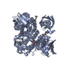
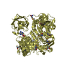
- PDB-7ccg: Crystal structure of ClA1, a kind of a chlorinase from soil bacteria -
+ Open data

- Basic information
| Entry | Database: PDB / ID: 7ccg | ||||||
|---|---|---|---|---|---|---|---|
| Title | Crystal structure of ClA1, a kind of a chlorinase from soil bacteria | ||||||
 Components Components | DNA-directed RNA polymerase subunit delta | ||||||
 Keywords Keywords | CELL INVASION / Chlorinated enzyme / ClA1 | ||||||
| Function / homology |  Function and homology information Function and homology information | ||||||
| Biological species |  Streptomyces albulus (bacteria) Streptomyces albulus (bacteria) | ||||||
| Method |  X-RAY DIFFRACTION / SYNCHROTRON / MOLECULAR REPLACEMENT / Resolution: 1.85 Å X-RAY DIFFRACTION / SYNCHROTRON / MOLECULAR REPLACEMENT / Resolution: 1.85 Å | ||||||
 Authors Authors | Ouyang, Z. / Li, Y. | ||||||
| Funding support | 1items
| ||||||
 Citation Citation | Journal: Biochem.Biophys.Res.Commun. / Year: 2020 Title: Crystal structure of ClA1, a type of chlorinase from soil bacteria. Authors: Miao, Y. / Yu, J. / Ouyang, Z. / Sun, H. / Li, Y. | ||||||
| History |
|
- Structure visualization
| Structure viewer | Molecule: MolmilJmol/JSmol |
|---|
- Downloads & links
-Download
| PDBx/mmCIF format | 7ccg.cif.gz | 132 KB | Display | PDBx/mmCIF format |
|---|---|---|---|---|
| PDB format | pdb7ccg.ent.gz | 83.6 KB | Display | PDB format |
| PDBx/mmJSON format | 7ccg.json.gz | Tree view | PDBx/mmJSON format | |
| Others |  Other downloads Other downloads |
-Validation report
| Arichive directory | https://data.pdbj.org/pub/pdb/validation_reports/cc/7ccgftp://data.pdbj.org/pub/pdb/validation_reports/cc/7ccg | HTTPS FTP |
|---|
-Related structure data
| Related structure data |  6rz2S S: Starting model for refinement |
|---|---|
| Similar structure data |


-Links
 PDBj
PDBj- Assembly
| Deposited unit | 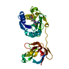
| ||||||||||||
|---|---|---|---|---|---|---|---|---|---|---|---|---|---|
| 1 | 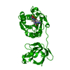
| ||||||||||||
| Unit cell |
| ||||||||||||
| Components on special symmetry positions |
|
-Components
| #1: Protein | Mass: 28291.092 Da / Num. of mol.: 1 Source method: isolated from a genetically manipulated source Source: (gene. exp.) Streptomyces albulus (bacteria) / Gene: DC74_7605, SALB_08549, SALB_08560 / Production host: |
|---|---|
| #2: Chemical | ChemComp-MET /   Type: L-peptide linking / Mass: 149.211 Da / Num. of mol.: 1 / Source method: obtained synthetically / Formula: C5H11NO2S / Feature type: SUBJECT OF INVESTIGATION Type: L-peptide linking / Mass: 149.211 Da / Num. of mol.: 1 / Source method: obtained synthetically / Formula: C5H11NO2S / Feature type: SUBJECT OF INVESTIGATION |
| #3: Chemical | ChemComp-5CD /   Mass: 285.687 Da / Num. of mol.: 1 / Source method: obtained synthetically / Formula: C10H12ClN5O3 / Feature type: SUBJECT OF INVESTIGATION Mass: 285.687 Da / Num. of mol.: 1 / Source method: obtained synthetically / Formula: C10H12ClN5O3 / Feature type: SUBJECT OF INVESTIGATION |
| #4: Water | ChemComp-HOH /  Mass: 18.015 Da / Num. of mol.: 87 / Source method: isolated from a natural source / Formula: H2O Mass: 18.015 Da / Num. of mol.: 87 / Source method: isolated from a natural source / Formula: H2O |
| Has ligand of interest | Y |
-Experimental details
-Experiment
| Experiment | Method: X-RAY DIFFRACTION / Number of used crystals: 1 |
|---|
- Sample preparation
| Crystal | Density Matthews: 2.41 Å3/Da / Density % sol: 48.93 % |
|---|---|
| Crystal grow | Temperature: 289.15 K / Method: evaporation Details: 0.1 M potassium chloride, 0.1 M sodium HEPES pH7.5, 15% w/v PEG 6000 |
-Data collection
| Diffraction | Mean temperature: 100 K / Serial crystal experiment: N |
|---|---|
| Diffraction source | Source: SYNCHROTRON / Site: SSRF  / Beamline: BL17U1 / Wavelength: 0.9791 Å / Beamline: BL17U1 / Wavelength: 0.9791 Å |
| Detector | Type: ADSC QUANTUM 315r / Detector: CCD / Date: Oct 7, 2019 |
| Radiation | Protocol: SINGLE WAVELENGTH / Monochromatic (M) / Laue (L): M / Scattering type: x-ray |
| Radiation wavelength | Wavelength: 0.9791 Å / Relative weight: 1 |
| Reflection | Resolution: 1.85→50 Å / Num. obs: 22423 / % possible obs: 99.6 % / Redundancy: 5.1 % / Biso Wilson estimate: 39.06 Å2 / CC1/2: 0.998 / Rrim(I) all: 0.046 / Net I/σ(I): 39.19 |
| Reflection shell | Resolution: 1.85→1.88 Å / Mean I/σ(I) obs: 1.555 / Num. unique obs: 1111 / CC1/2: 0.76 / Rrim(I) all: 0.854 |
- Processing
| Software |
| |||||||||||||||||||||||||||||||||||||||||||||||||||||||||||||||||||||||||||||||||||||||||||||||||||||||||
|---|---|---|---|---|---|---|---|---|---|---|---|---|---|---|---|---|---|---|---|---|---|---|---|---|---|---|---|---|---|---|---|---|---|---|---|---|---|---|---|---|---|---|---|---|---|---|---|---|---|---|---|---|---|---|---|---|---|---|---|---|---|---|---|---|---|---|---|---|---|---|---|---|---|---|---|---|---|---|---|---|---|---|---|---|---|---|---|---|---|---|---|---|---|---|---|---|---|---|---|---|---|---|---|---|---|---|
| Refinement | Method to determine structure: MOLECULAR REPLACEMENT Starting model: 6RZ2 Resolution: 1.85→5 Å / SU ML: 0.2253 / Cross valid method: FREE R-VALUE / σ(F): 2.02 / Phase error: 26.7583 Stereochemistry target values: GeoStd + Monomer Library + CDL v1.2
| |||||||||||||||||||||||||||||||||||||||||||||||||||||||||||||||||||||||||||||||||||||||||||||||||||||||||
| Solvent computation | Shrinkage radii: 0.9 Å / VDW probe radii: 1.11 Å / Solvent model: FLAT BULK SOLVENT MODEL | |||||||||||||||||||||||||||||||||||||||||||||||||||||||||||||||||||||||||||||||||||||||||||||||||||||||||
| Displacement parameters | Biso mean: 48.07 Å2 | |||||||||||||||||||||||||||||||||||||||||||||||||||||||||||||||||||||||||||||||||||||||||||||||||||||||||
| Refinement step | Cycle: LAST / Resolution: 1.85→5 Å
| |||||||||||||||||||||||||||||||||||||||||||||||||||||||||||||||||||||||||||||||||||||||||||||||||||||||||
| Refine LS restraints |
| |||||||||||||||||||||||||||||||||||||||||||||||||||||||||||||||||||||||||||||||||||||||||||||||||||||||||
| LS refinement shell |
|
