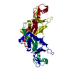
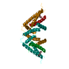
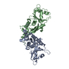
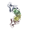
[English] 日本語
Yorodumi
- PDB-4zud: Crystal Structure of Human Angiotensin Receptor in Complex with I... -

+
Open data


ID or keywords:

Loading...

-
Basic information

Entry
Database: PDB / ID: 4zud
TitleCrystal Structure of Human Angiotensin Receptor in Complex with Inverse Agonist Olmesartan at 2.8A resolution.
ComponentsChimera protein of Soluble cytochrome b562 and Type-1 angiotensin II receptor
KeywordsMEMBRANE PROTEIN / human Angiotensin Receptor AT1R / BRIL / G Protein-Coupled Receptor / GPCR / GPCR network / Lipidic Cubic Phase / LCP / structural genomics / olmesartan / angiotensin receptor blocker / anti-hypertensive drug / PSI-Biology / SIGNALING PROTEIN
Function / homology
Function and homology information


angiotensin type I receptor activity / positive regulation of phospholipase A2 activity / angiotensin type II receptor activity / phospholipase C-activating angiotensin-activated signaling pathway / regulation of renal sodium excretion / maintenance of blood vessel diameter homeostasis by renin-angiotensin / bradykinin receptor binding / renin-angiotensin regulation of aldosterone production / positive regulation of blood vessel endothelial cell proliferation involved in sprouting angiogenesis / positive regulation of macrophage derived foam cell differentiation ...angiotensin type I receptor activity / positive regulation of phospholipase A2 activity / angiotensin type II receptor activity / phospholipase C-activating angiotensin-activated signaling pathway / regulation of renal sodium excretion / maintenance of blood vessel diameter homeostasis by renin-angiotensin / bradykinin receptor binding / renin-angiotensin regulation of aldosterone production / positive regulation of blood vessel endothelial cell proliferation involved in sprouting angiogenesis / positive regulation of macrophage derived foam cell differentiation / C-C chemokine receptor activity / C-C chemokine binding / positive regulation of CoA-transferase activity / low-density lipoprotein particle remodeling / regulation of systemic arterial blood pressure by renin-angiotensin / Rho protein signal transduction / regulation of vasoconstriction / positive regulation of protein metabolic process / blood vessel diameter maintenance / Peptide ligand-binding receptors / neurogenesis / cell chemotaxis / kidney development / angiotensin-activated signaling pathway / regulation of cell growth / calcium-mediated signaling / electron transport chain / brain development / positive regulation of inflammatory response / positive regulation of reactive oxygen species metabolic process / Cargo recognition for clathrin-mediated endocytosis / Clathrin-mediated endocytosis / phospholipase C-activating G protein-coupled receptor signaling pathway / regulation of cell population proliferation / positive regulation of cytosolic calcium ion concentration / regulation of inflammatory response / G alpha (q) signalling events / periplasmic space / electron transfer activity / inflammatory response / symbiont entry into host cell / iron ion binding / immune response / G protein-coupled receptor signaling pathway / protein heterodimerization activity / external side of plasma membrane / heme binding / membrane / plasma membrane
Similarity search - Function
Angiotensin II receptor type 1 / Angiotensin II receptor family / Cytochrome b562 / Cytochrome b562 / Cytochrome c/b562 / Serpentine type 7TM GPCR chemoreceptor Srsx / G-protein coupled receptors family 1 signature. / G protein-coupled receptor, rhodopsin-like / GPCR, rhodopsin-like, 7TM / G-protein coupled receptors family 1 profile. / 7 transmembrane receptor (rhodopsin family)
Similarity search - Domain/homology
Olmesartan / Soluble cytochrome b562 / Type-1 angiotensin II receptor
Similarity search - Component
Biological speciesEscherichia coli (E. coli)
Homo sapiens (human)
MethodX-RAY DIFFRACTION / SYNCHROTRON / MOLECULAR REPLACEMENT / Resolution: 2.8 Å
AuthorsZhang, H. / Unal, H. / Desnoyer, R. / Han, G.W. / Patel, N. / Katritch, V. / Karnik, S.S. / Cherezov, V. / Stevens, R.C. / GPCR Network (GPCR)
Funding support United States, 1items
OrganizationGrant numberCountry
National Institutes of Health/National Institute of General Medical Sciences (NIH/NIGMS)U54 GM094618 United States
CitationJournal: J.Biol.Chem. / Year: 2015
Title: Structural Basis for Ligand Recognition and Functional Selectivity at Angiotensin Receptor.
Authors: Zhang, H. / Unal, H. / Desnoyer, R. / Han, G.W. / Patel, N. / Katritch, V. / Karnik, S.S. / Cherezov, V. / Stevens, R.C.
History
DepositionMay 15, 2015Deposition site: RCSB / Processing site: RCSB
Revision 1.0Oct 7, 2015Provider: repository / Type: Initial release
Revision 1.1Oct 14, 2015Group: Database references
Revision 1.2Dec 16, 2015Group: Database references
Revision 1.3Sep 20, 2017Group: Author supporting evidence / Database references / Derived calculations
Category: citation / pdbx_audit_support / pdbx_struct_oper_list
Item: _citation.journal_id_CSD / _pdbx_audit_support.funding_organization / _pdbx_struct_oper_list.symmetry_operation
Revision 1.4Dec 25, 2019Group: Author supporting evidence / Category: pdbx_audit_support / Item: _pdbx_audit_support.funding_organization
Revision 1.5Sep 27, 2023Group: Data collection / Database references / Refinement description
Category: chem_comp_atom / chem_comp_bond ...chem_comp_atom / chem_comp_bond / database_2 / pdbx_initial_refinement_model
Item: _database_2.pdbx_DOI / _database_2.pdbx_database_accession

-
Structure visualization

Structure viewerMolecule:
MolmilJmol/JSmol

Downloads & links

-
Assembly

Deposited unit
A: Chimera protein of Soluble cytochrome b562 and Type-1 angiotensin II receptor
hetero molecules


Theoretical massNumber of molelcules
Total (without water)47,1692
Polymers46,7221
Non-polymers4471
Water00
1


  • Idetical with deposited unit
  • defined by software
TypeNameSymmetry operationNumber
identity operation1_555x,y,z1
MethodPISA
Unit cell
Length a, b, c (Å)41.200, 41.200, 251.160
Angle α, β, γ (deg.)90.00, 90.00, 120.00
Int Tables number145
Space group name H-MP32
DetailsAuthors state that the biological unit is unknown

-
Components

#1: Protein Chimera protein of Soluble cytochrome b562 and Type-1 angiotensin II receptor / Cytochrome b-562 / AT1AR / AT1BR / Angiotensin II type-1 receptor / AT1 / AT1AR / AT1BR / ...Cytochrome b-562 / AT1AR / AT1BR / Angiotensin II type-1 receptor / AT1 / AT1AR / AT1BR / Angiotensin II type-1 receptor / AT1


Mass: 46722.043 Da / Num. of mol.: 1 / Mutation: M1007W, H1102I, R1106L
Source method: isolated from a genetically manipulated source
Source: (gene. exp.) Escherichia coli (E. coli), (gene. exp.) Homo sapiens (human)
Gene: cybC, AGTR1, AGTR1A, AGTR1B, AT2R1, AT2R1B / Plasmid: pFASTBAC / Cell line (production host): sf9 / Production host: Spodoptera frugiperda (fall armyworm) / Strain (production host): sf9 / References: UniProt: P0ABE7, UniProt: P30556
#2: Chemical ChemComp-OLM / Olmesartan


Mass: 446.502 Da / Num. of mol.: 1 / Source method: obtained synthetically / Formula: C24H26N6O3 / Comment: medication*YM

-
Experimental details

-
Experiment

ExperimentMethod: X-RAY DIFFRACTION

-
Sample preparation

CrystalDensity Matthews: 2.9 Å3/Da / Density % sol: 57.54 %
Crystal growTemperature: 293 K / Method: lipidic cubic phase / pH: 5
Details: 100 mM sodium citrate, pH 5.0, 400 mM KH2PO4, 25% (v/v) PEG400, and 6% (v/v) DMSO

-
Data collection

DiffractionMean temperature: 100 K
Diffraction sourceSource: SYNCHROTRON / Site: APS / Beamline: 23-ID-D / Wavelength: 1.033 Å
DetectorType: PSI PILATUS 6M / Detector: PIXEL / Date: Dec 31, 2014
RadiationProtocol: SINGLE WAVELENGTH / Monochromatic (M) / Laue (L): M / Scattering type: x-ray
Radiation wavelengthWavelength: 1.033 Å / Relative weight: 1
Reflection twin
Crystal-IDIDOperatorDomain-IDFraction
11H, K, L10.575
11K, H, -L20.425
ReflectionResolution: 2.8→35.68 Å / Num. obs: 11028 / % possible obs: 94 % / Redundancy: 2.36 % / Rmerge(I) obs: 0.054 / Net I/σ(I): 11.05
Reflection shellResolution: 2.8→2.87 Å / Redundancy: 2.09 % / Rmerge(I) obs: 0.758 / Mean I/σ(I) obs: 1.17 / % possible all: 85.3

-
Processing

Software
NameVersionClassification
REFMAC5.8.0103refinement
XDSdata reduction
XSCALEdata scaling
PHASERphasing
RefinementMethod to determine structure: MOLECULAR REPLACEMENT
Starting model: PDB 4YAY
Resolution: 2.8→35.68 Å / Cor.coef. Fo:Fc: 0.948 / Cor.coef. Fo:Fc free: 0.904 / SU B: 20.583 / SU ML: 0.21 / Cross valid method: THROUGHOUT / ESU R Free: 0.077 / Stereochemistry target values: MAXIMUM LIKELIHOOD / Details: HYDROGENS HAVE BEEN ADDED IN THE RIDING POSITIONS
RfactorNum. reflection% reflectionSelection details
Rfree0.2345 555 5 %RANDOM
Rwork0.19383 ---
obs0.19588 10472 93.96 %-
Solvent computationIon probe radii: 0.8 Å / Shrinkage radii: 0.8 Å / VDW probe radii: 1.2 Å / Solvent model: MASK
Displacement parametersBiso mean: 84.956 Å2
Baniso -1Baniso -2Baniso -3
1-28.35 Å20 Å20 Å2
2--28.35 Å20 Å2
3----56.69 Å2
Refinement stepCycle: LAST / Resolution: 2.8→35.68 Å
ProteinNucleic acidLigandSolventTotal
Num. atoms2918 0 33 0 2951
Refine LS restraints
Refine-IDTypeDev idealDev ideal targetNumber
X-RAY DIFFRACTIONr_bond_refined_d0.0090.023021
X-RAY DIFFRACTIONr_bond_other_d0.0020.022941
X-RAY DIFFRACTIONr_angle_refined_deg1.1331.9774123
X-RAY DIFFRACTIONr_angle_other_deg0.9593.0026718
X-RAY DIFFRACTIONr_dihedral_angle_1_deg5.9095372
X-RAY DIFFRACTIONr_dihedral_angle_2_deg36.19324.123114
X-RAY DIFFRACTIONr_dihedral_angle_3_deg15.16515483
X-RAY DIFFRACTIONr_dihedral_angle_4_deg11.716159
X-RAY DIFFRACTIONr_chiral_restr0.0590.2496
X-RAY DIFFRACTIONr_gen_planes_refined0.0050.0213342
X-RAY DIFFRACTIONr_gen_planes_other0.0010.02687
X-RAY DIFFRACTIONr_nbd_refined
X-RAY DIFFRACTIONr_nbd_other
X-RAY DIFFRACTIONr_nbtor_refined
X-RAY DIFFRACTIONr_nbtor_other
X-RAY DIFFRACTIONr_xyhbond_nbd_refined
X-RAY DIFFRACTIONr_xyhbond_nbd_other
X-RAY DIFFRACTIONr_metal_ion_refined
X-RAY DIFFRACTIONr_metal_ion_other
X-RAY DIFFRACTIONr_symmetry_vdw_refined
X-RAY DIFFRACTIONr_symmetry_vdw_other
X-RAY DIFFRACTIONr_symmetry_hbond_refined
X-RAY DIFFRACTIONr_symmetry_hbond_other
X-RAY DIFFRACTIONr_symmetry_metal_ion_refined
X-RAY DIFFRACTIONr_symmetry_metal_ion_other
X-RAY DIFFRACTIONr_mcbond_it1.5257.6151500
X-RAY DIFFRACTIONr_mcbond_other1.5257.6161499
X-RAY DIFFRACTIONr_mcangle_it2.65811.4161868
X-RAY DIFFRACTIONr_mcangle_other2.65711.4151869
X-RAY DIFFRACTIONr_scbond_it1.0877.5691521
X-RAY DIFFRACTIONr_scbond_other1.0877.5691522
X-RAY DIFFRACTIONr_scangle_it
X-RAY DIFFRACTIONr_scangle_other1.89511.3332256
X-RAY DIFFRACTIONr_long_range_B_refined4.43161.0623512
X-RAY DIFFRACTIONr_long_range_B_other4.4361.0653513
X-RAY DIFFRACTIONr_rigid_bond_restr
X-RAY DIFFRACTIONr_sphericity_free
X-RAY DIFFRACTIONr_sphericity_bonded
LS refinement shellResolution: 2.8→2.873 Å / Total num. of bins used: 20
RfactorNum. reflection% reflection
Rfree0.275 32 -
Rwork0.214 699 -
obs--85 %
Refinement TLS params.

Method: refined / Refine-ID: X-RAY DIFFRACTION

IDL112)L122)L132)L222)L232)L332)S11 (Å °)S12 (Å °)S13 (Å °)S21 (Å °)S22 (Å °)S23 (Å °)S31 (Å °)S32 (Å °)S33 (Å °)T112)T122)T132)T222)T232)T332)Origin x (Å)Origin y (Å)Origin z (Å)
10.0882-0.24080.322.3768-1.17851.3014-0.04160.0249-0.00360.0387-0.0445-0.1056-0.13490.03660.08610.03220.0020.01220.10810.01470.1438-23.160872.68781.7663
20.58990.0982-0.01890.05010.13411.33070.0267-0.09720.0322-0.0021-0.0328-0.01510.12260.10410.00610.03910.01570.03720.1011-0.03740.1645-40.615263.657136.7014
Refinement TLS group
IDRefine-IDRefine TLS-IDSelection detailsAuth asym-IDAuth seq-ID
1X-RAY DIFFRACTION1{ A | 1002 - 1106 }A1002 - 1106
2X-RAY DIFFRACTION2{ A | 12 - 304 }A12 - 304

+
About Yorodumi

-
News

-
Feb 9, 2022. New format data for meta-information of EMDB entries

New format data for meta-information of EMDB entries

  • Version 3 of the EMDB header file is now the official format.
  • The previous official version 1.9 will be removed from the archive.

Related info.:EMDB header

External links:wwPDB to switch to version 3 of the EMDB data model

-
Aug 12, 2020. Covid-19 info

Covid-19 info

URL: https://pdbjlvh1.pdbj.org/emnavi/covid19.php

New page: Covid-19 featured information page in EM Navigator.

Related info.:Covid-19 info / Mar 5, 2020. Novel coronavirus structure data

+
Mar 5, 2020. Novel coronavirus structure data

Novel coronavirus structure data

Related info.:Yorodumi Speices / Aug 12, 2020. Covid-19 info

External links:COVID-19 featured content - PDBj / Molecule of the Month (242):Coronavirus Proteases

+
Jan 31, 2019. EMDB accession codes are about to change! (news from PDBe EMDB page)

EMDB accession codes are about to change! (news from PDBe EMDB page)

  • The allocation of 4 digits for EMDB accession codes will soon come to an end. Whilst these codes will remain in use, new EMDB accession codes will include an additional digit and will expand incrementally as the available range of codes is exhausted. The current 4-digit format prefixed with “EMD-” (i.e. EMD-XXXX) will advance to a 5-digit format (i.e. EMD-XXXXX), and so on. It is currently estimated that the 4-digit codes will be depleted around Spring 2019, at which point the 5-digit format will come into force.
  • The EM Navigator/Yorodumi systems omit the EMD- prefix.

Related info.:Q: What is EMD? / ID/Accession-code notation in Yorodumi/EM Navigator

External links:EMDB Accession Codes are Changing Soon! / Contact to PDBj

+
Jul 12, 2017. Major update of PDB

Major update of PDB

  • wwPDB released updated PDB data conforming to the new PDBx/mmCIF dictionary.
  • This is a major update changing the version number from 4 to 5, and with Remediation, in which all the entries are updated.
  • In this update, many items about electron microscopy experimental information are reorganized (e.g. em_software).
  • Now, EM Navigator and Yorodumi are based on the updated data.

External links:wwPDB Remediation / Enriched Model Files Conforming to OneDep Data Standards Now Available in the PDB FTP Archive

-
Yorodumi

Thousand views of thousand structures

  • Yorodumi is a browser for structure data from EMDB, PDB, SASBDB, etc.
  • This page is also the successor to EM Navigator detail page, and also detail information page/front-end page for Omokage search.
  • The word "yorodu" (or yorozu) is an old Japanese word meaning "ten thousand". "mi" (miru) is to see.

Related info.:EMDB / PDB / SASBDB / Comparison of 3 databanks / Yorodumi Search / Aug 31, 2016. New EM Navigator & Yorodumi / Yorodumi Papers / Jmol/JSmol / Function and homology information / Changes in new EM Navigator and Yorodumi

Read more