 Movie
Movie Controller

[English] 日本語
 Yorodumi
- PDB-4zi5: Crystal Structure of Dienelactone Hydrolase-like Promiscuous Phos... -
+ Open data

- Basic information
| Entry | Database: PDB / ID: 4zi5 | ||||||
|---|---|---|---|---|---|---|---|
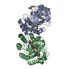
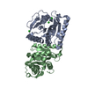
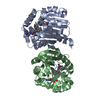
| Title | Crystal Structure of Dienelactone Hydrolase-like Promiscuous Phospotriesterase P91 from Metagenomic Libraries | ||||||
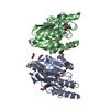
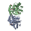
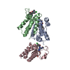
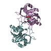
 Components Components | P91 | ||||||
 Keywords Keywords | HYDROLASE / Metagenomic Libraries / alpha/beta Hydrolase / Promiscuity / Phosphotriesterase | ||||||
| Function / homology | Alpha/Beta hydrolase fold, catalytic domain / Rossmann fold / 3-Layer(aba) Sandwich / Alpha Beta Function and homology information Function and homology information | ||||||
| Biological species | metagenome (others) | ||||||
| Method |  X-RAY DIFFRACTION / SYNCHROTRON / MOLECULAR REPLACEMENT / Resolution: 1.702 Å X-RAY DIFFRACTION / SYNCHROTRON / MOLECULAR REPLACEMENT / Resolution: 1.702 Å | ||||||
 Authors Authors | Colin, P.-Y. / Fischer, G. / Hyvonen, M. / Hollfelder, F. | ||||||
 Citation Citation | Journal: Nat Commun / Year: 2015 Title: Ultrahigh-throughput discovery of promiscuous enzymes by picodroplet functional metagenomics. Authors: Colin, P.Y. / Kintses, B. / Gielen, F. / Miton, C.M. / Fischer, G. / Mohamed, M.F. / Hyvonen, M. / Morgavi, D.P. / Janssen, D.B. / Hollfelder, F. | ||||||
| History |
|
- Structure visualization
| Structure viewer | Molecule: MolmilJmol/JSmol |
|---|
- Downloads & links
-Download
| PDBx/mmCIF format | 4zi5.cif.gz | 114.3 KB | Display | PDBx/mmCIF format |
|---|---|---|---|---|
| PDB format | pdb4zi5.ent.gz | 87.1 KB | Display | PDB format |
| PDBx/mmJSON format | 4zi5.json.gz | Tree view | PDBx/mmJSON format | |
| Others |  Other downloads Other downloads |
-Validation report
| Arichive directory | https://data.pdbj.org/pub/pdb/validation_reports/zi/4zi5ftp://data.pdbj.org/pub/pdb/validation_reports/zi/4zi5 | HTTPS FTP |
|---|
-Related structure data
| Related structure data |  3f67S S: Starting model for refinement |
|---|---|
| Similar structure data |


-Links
 PDBj
PDBj- Assembly
| Deposited unit | 
| ||||||||||||||||||
|---|---|---|---|---|---|---|---|---|---|---|---|---|---|---|---|---|---|---|---|
| 1 |
| ||||||||||||||||||
| Unit cell |
| ||||||||||||||||||
| Noncrystallographic symmetry (NCS) | NCS domain:
NCS domain segments: Component-ID: _ / Ens-ID: 1 / Beg auth comp-ID: GLY / Beg label comp-ID: GLY / End auth comp-ID: GLY / End label comp-ID: GLY / Refine code: _ / Auth seq-ID: -1 - 237 / Label seq-ID: 12 - 250
|
-Components
| #1: Protein | Mass: 26867.133 Da / Num. of mol.: 2 Source method: isolated from a genetically manipulated source Source: (gene. exp.) metagenome (others) / Plasmid: pASKIBA5+ / Production host:  #2: Chemical |   Mass: 24.305 Da / Num. of mol.: 2 / Source method: obtained synthetically / Formula: Mg Mass: 24.305 Da / Num. of mol.: 2 / Source method: obtained synthetically / Formula: Mg#3: Chemical | ChemComp-CL / |   Mass: 35.453 Da / Num. of mol.: 1 / Source method: obtained synthetically / Formula: Cl Mass: 35.453 Da / Num. of mol.: 1 / Source method: obtained synthetically / Formula: Cl#4: Water | ChemComp-HOH / |  Mass: 18.015 Da / Num. of mol.: 456 / Source method: isolated from a natural source / Formula: H2O Mass: 18.015 Da / Num. of mol.: 456 / Source method: isolated from a natural source / Formula: H2O |
|---|
-Experimental details
-Experiment
| Experiment | Method: X-RAY DIFFRACTION |
|---|
- Sample preparation
| Crystal | Density Matthews: 2.26 Å3/Da / Density % sol: 45.56 % / Description: shard-like plates |
|---|---|
| Crystal grow | Temperature: 292 K / Method: vapor diffusion, sitting drop / pH: 8.5 / Details: 0.1M Tris pH=8.5, 0.2M MgCl2, 20% PEG8000 |
-Data collection
| Diffraction | Mean temperature: 100 K |
|---|---|
| Diffraction source | Source: SYNCHROTRON / Site: SLS  / Beamline: X06DA / Wavelength: 0.91882 Å / Beamline: X06DA / Wavelength: 0.91882 Å |
| Detector | Type: DECTRIS PILATUS 2M / Detector: PIXEL / Date: Jun 29, 2014 |
| Radiation | Protocol: SINGLE WAVELENGTH / Monochromatic (M) / Laue (L): M / Scattering type: x-ray |
| Radiation wavelength | Wavelength: 0.91882 Å / Relative weight: 1 |
| Reflection | Resolution: 1.702→61.57 Å / Num. obs: 45156 / % possible obs: 87.6 % / Observed criterion σ(I): -3 / Redundancy: 3.8 % / Biso Wilson estimate: 18.4 Å2 / Rmerge(I) obs: 0.063 / Rsym value: 0.063 / Net I/σ(I): 12.5 |
| Reflection shell | Resolution: 1.702→1.708 Å / Redundancy: 2.8 % / Rmerge(I) obs: 0.454 / Mean I/σ(I) obs: 2 / Rsym value: 0.454 / % possible all: 55.6 |
- Processing
| Software |
| |||||||||||||||||||||
|---|---|---|---|---|---|---|---|---|---|---|---|---|---|---|---|---|---|---|---|---|---|---|
| Refinement | Method to determine structure: MOLECULAR REPLACEMENT Starting model: 3F67 Resolution: 1.702→61.57 Å / Cor.coef. Fo:Fc: 0.967 / Cor.coef. Fo:Fc free: 0.963 / SU B: 2.654 / SU ML: 0.082 / Cross valid method: THROUGHOUT / ESU R: 0.131 / ESU R Free: 0.117 / Stereochemistry target values: MAXIMUM LIKELIHOOD / Details: HYDROGENS HAVE BEEN ADDED IN THE RIDING POSITIONS
| |||||||||||||||||||||
| Solvent computation | Ion probe radii: 0.8 Å / Shrinkage radii: 0.8 Å / VDW probe radii: 1.2 Å / Solvent model: MASK | |||||||||||||||||||||
| Displacement parameters | Biso mean: 20.248 Å2
| |||||||||||||||||||||
| Refinement step | Cycle: LAST / Resolution: 1.702→61.57 Å
| |||||||||||||||||||||
| Refine LS restraints NCS | Ens-ID: 1 / Number: 28118 / Refine-ID: X-RAY DIFFRACTION / Type: interatomic distance / Rms dev position: 0.06 Å / Weight position: 0.05
| |||||||||||||||||||||
| LS refinement shell | Resolution: 1.702→1.746 Å / Total num. of bins used: 20
|
