 Movie
Movie Controller

[English] 日本語
 Yorodumi
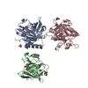
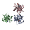
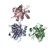
- PDB-4zfx: Siderocalin-mediated recognition and cellular uptake of actinides -
+ Open data

- Basic information
| Entry | Database: PDB / ID: 4zfx | ||||||
|---|---|---|---|---|---|---|---|
| Title | Siderocalin-mediated recognition and cellular uptake of actinides | ||||||
 Components Components | Neutrophil gelatinase-associated lipocalin | ||||||
 Keywords Keywords | Metal Binding Protein/inhibitor / Metal Binding Protein-inhibitor complex | ||||||
| Function / homology |  Function and homology information Function and homology informationpositive regulation of iron ion import across plasma membrane / positive regulation of hippocampal neuron apoptotic process / positive regulation of endothelial tube morphogenesis / negative regulation of hippocampal neuron apoptotic process / positive regulation of cell projection organization / Metal sequestration by antimicrobial proteins / response to mycotoxin / response to kainic acid / siderophore transport / cellular response to increased oxygen levels ...positive regulation of iron ion import across plasma membrane / positive regulation of hippocampal neuron apoptotic process / positive regulation of endothelial tube morphogenesis / negative regulation of hippocampal neuron apoptotic process / positive regulation of cell projection organization / Metal sequestration by antimicrobial proteins / response to mycotoxin / response to kainic acid / siderophore transport / cellular response to increased oxygen levels / response to blue light / cellular response to X-ray / response to fructose / short-term memory / cellular response to interleukin-6 / enterobactin binding / iron ion sequestering activity / response to herbicide / response to iron(II) ion / positive regulation of reactive oxygen species biosynthetic process / cellular response to interleukin-1 / long-term memory / extrinsic apoptotic signaling pathway in absence of ligand / positive regulation of endothelial cell migration / cellular response to nutrient levels / acute-phase response / cellular response to nerve growth factor stimulus / Iron uptake and transport / specific granule lumen / cellular response to tumor necrosis factor / cellular response to amyloid-beta / response to virus / cellular response to hydrogen peroxide / positive regulation of cold-induced thermogenesis / cellular response to lipopolysaccharide / protease binding / Interleukin-4 and Interleukin-13 signaling / cellular response to hypoxia / defense response to bacterium / iron ion binding / response to xenobiotic stimulus / innate immune response / Neutrophil degranulation / positive regulation of gene expression / : / extracellular exosome / extracellular region / identical protein binding Similarity search - Function | ||||||
| Biological species |  Homo sapiens (human) Homo sapiens (human) | ||||||
| Method |  X-RAY DIFFRACTION / MOLECULAR REPLACEMENT / Resolution: 2.55 Å X-RAY DIFFRACTION / MOLECULAR REPLACEMENT / Resolution: 2.55 Å | ||||||
 Authors Authors | Allred, B.E. / Rupert, P.B. / Gauny, S.S. / An, D.D. / Ralston, C.Y. / Sturzbecher-Hoehne, M. / Strong, R.K. / Abergel, R.J. | ||||||
 Citation Citation | Journal: Proc.Natl.Acad.Sci.USA / Year: 2015 Title: Siderocalin-mediated recognition, sensitization, and cellular uptake of actinides. Authors: Allred, B.E. / Rupert, P.B. / Gauny, S.S. / An, D.D. / Ralston, C.Y. / Sturzbecher-Hoehne, M. / Strong, R.K. / Abergel, R.J. | ||||||
| History |
|
- Structure visualization
| Structure viewer | Molecule: MolmilJmol/JSmol |
|---|
- Downloads & links
-Download
| PDBx/mmCIF format | 4zfx.cif.gz | 223.4 KB | Display | PDBx/mmCIF format |
|---|---|---|---|---|
| PDB format | pdb4zfx.ent.gz | 180.1 KB | Display | PDB format |
| PDBx/mmJSON format | 4zfx.json.gz | Tree view | PDBx/mmJSON format | |
| Others |  Other downloads Other downloads |
-Validation report
| Arichive directory | https://data.pdbj.org/pub/pdb/validation_reports/zf/4zfxftp://data.pdbj.org/pub/pdb/validation_reports/zf/4zfx | HTTPS FTP |
|---|
-Related structure data
| Related structure data |  4zhcC  4zhdC  4zhfC  4zhgC  4zhhC 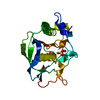 1lm6S S: Starting model for refinement C: citing same article ( |
|---|---|
| Similar structure data |


-Links
 PDBj
PDBj

- Assembly
| Deposited unit | 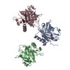
| |||||||||||||||||||||||||||||||||||||||||||||||||||||||||||||||||||||||||||||||||
|---|---|---|---|---|---|---|---|---|---|---|---|---|---|---|---|---|---|---|---|---|---|---|---|---|---|---|---|---|---|---|---|---|---|---|---|---|---|---|---|---|---|---|---|---|---|---|---|---|---|---|---|---|---|---|---|---|---|---|---|---|---|---|---|---|---|---|---|---|---|---|---|---|---|---|---|---|---|---|---|---|---|---|
| 1 |
| |||||||||||||||||||||||||||||||||||||||||||||||||||||||||||||||||||||||||||||||||
| Unit cell |
| |||||||||||||||||||||||||||||||||||||||||||||||||||||||||||||||||||||||||||||||||
| Noncrystallographic symmetry (NCS) | NCS domain:
NCS domain segments: Component-ID: _ / End auth comp-ID: ILE / End label comp-ID: ILE / Refine code: _
NCS ensembles :
|
-Components
| #1: Protein | Mass: 20700.564 Da / Num. of mol.: 3 / Fragment: residues 24-197 Source method: isolated from a genetically manipulated source Source: (gene. exp.) Homo sapiens (human) / Gene: LCN2, HNL, NGAL / Plasmid: pGEX / Production host:  #2: Chemical |   Mass: 232.038 Da / Num. of mol.: 3 / Source method: obtained synthetically / Formula: Th Mass: 232.038 Da / Num. of mol.: 3 / Source method: obtained synthetically / Formula: Th#3: Chemical | 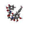  Mass: 669.546 Da / Num. of mol.: 2 / Source method: obtained synthetically / Formula: C30H27N3O15 Mass: 669.546 Da / Num. of mol.: 2 / Source method: obtained synthetically / Formula: C30H27N3O15#4: Chemical | ChemComp-SO4 / |   Mass: 96.063 Da / Num. of mol.: 1 / Source method: obtained synthetically / Formula: SO4 Mass: 96.063 Da / Num. of mol.: 1 / Source method: obtained synthetically / Formula: SO4#5: Water | ChemComp-HOH / |  Mass: 18.015 Da / Num. of mol.: 21 / Source method: isolated from a natural source / Formula: H2O Mass: 18.015 Da / Num. of mol.: 21 / Source method: isolated from a natural source / Formula: H2OHas protein modification | Y | |
|---|
-Experimental details
-Experiment
| Experiment | Method: X-RAY DIFFRACTION / Number of used crystals: 1 |
|---|
- Sample preparation
| Crystal | Density Matthews: 3.24 Å3/Da / Density % sol: 62.02 % |
|---|---|
| Crystal grow | Temperature: 298 K / Method: vapor diffusion / pH: 4 / Details: NaCl, Li2SO4, Acetate, Ammonium Sulfate |
-Data collection
| Diffraction | Mean temperature: 100 K | ||||||||||||||||||||||||||||||||||||||||||||||||||||||||||||||||||||||||||||||||||||||||||||||||||||||||||||||||||||||||||||||
|---|---|---|---|---|---|---|---|---|---|---|---|---|---|---|---|---|---|---|---|---|---|---|---|---|---|---|---|---|---|---|---|---|---|---|---|---|---|---|---|---|---|---|---|---|---|---|---|---|---|---|---|---|---|---|---|---|---|---|---|---|---|---|---|---|---|---|---|---|---|---|---|---|---|---|---|---|---|---|---|---|---|---|---|---|---|---|---|---|---|---|---|---|---|---|---|---|---|---|---|---|---|---|---|---|---|---|---|---|---|---|---|---|---|---|---|---|---|---|---|---|---|---|---|---|---|---|---|
| Diffraction source | Source: ROTATING ANODE / Type: RIGAKU MICROMAX-007 HF / Wavelength: 1.54 Å | ||||||||||||||||||||||||||||||||||||||||||||||||||||||||||||||||||||||||||||||||||||||||||||||||||||||||||||||||||||||||||||||
| Detector | Type: RIGAKU SATURN 944+ / Detector: CCD / Date: Mar 13, 2014 | ||||||||||||||||||||||||||||||||||||||||||||||||||||||||||||||||||||||||||||||||||||||||||||||||||||||||||||||||||||||||||||||
| Radiation | Monochromator: VariMax HF mirrors / Protocol: SINGLE WAVELENGTH / Monochromatic (M) / Laue (L): M / Scattering type: x-ray | ||||||||||||||||||||||||||||||||||||||||||||||||||||||||||||||||||||||||||||||||||||||||||||||||||||||||||||||||||||||||||||||
| Radiation wavelength | Wavelength: 1.54 Å / Relative weight: 1 | ||||||||||||||||||||||||||||||||||||||||||||||||||||||||||||||||||||||||||||||||||||||||||||||||||||||||||||||||||||||||||||||
| Reflection | Resolution: 2.55→50 Å / Num. obs: 26474 / % possible obs: 99.7 % / Redundancy: 7.6 % / Rmerge(I) obs: 0.082 / Χ2: 1.883 / Net I/av σ(I): 27.788 / Net I/σ(I): 17.5 / Num. measured all: 200751 | ||||||||||||||||||||||||||||||||||||||||||||||||||||||||||||||||||||||||||||||||||||||||||||||||||||||||||||||||||||||||||||||
| Reflection shell | Diffraction-ID: 1 / Rejects: _
|
- Processing
| Software |
| |||||||||||||||||||||||||||||||||||||||||||||||||||||||||||||||||||||||||||||||||||||||||||||||||||||||||||||||||||||||||||||||||||||||||||||||||||||||||||||||||||||||||||||||
|---|---|---|---|---|---|---|---|---|---|---|---|---|---|---|---|---|---|---|---|---|---|---|---|---|---|---|---|---|---|---|---|---|---|---|---|---|---|---|---|---|---|---|---|---|---|---|---|---|---|---|---|---|---|---|---|---|---|---|---|---|---|---|---|---|---|---|---|---|---|---|---|---|---|---|---|---|---|---|---|---|---|---|---|---|---|---|---|---|---|---|---|---|---|---|---|---|---|---|---|---|---|---|---|---|---|---|---|---|---|---|---|---|---|---|---|---|---|---|---|---|---|---|---|---|---|---|---|---|---|---|---|---|---|---|---|---|---|---|---|---|---|---|---|---|---|---|---|---|---|---|---|---|---|---|---|---|---|---|---|---|---|---|---|---|---|---|---|---|---|---|---|---|---|---|---|---|
| Refinement | Method to determine structure: MOLECULAR REPLACEMENT Starting model: 1LM6 Resolution: 2.55→50 Å / Cor.coef. Fo:Fc: 0.92 / Cor.coef. Fo:Fc free: 0.911 / WRfactor Rfree: 0.2619 / WRfactor Rwork: 0.2277 / FOM work R set: 0.8 / SU B: 16.495 / SU ML: 0.2 / SU R Cruickshank DPI: 0.4516 / SU Rfree: 0.287 / Cross valid method: THROUGHOUT / σ(F): 0 / ESU R: 0.452 / ESU R Free: 0.287 / Stereochemistry target values: MAXIMUM LIKELIHOOD Details: HYDROGENS HAVE BEEN ADDED IN THE RIDING POSITIONS U VALUES : WITH TLS ADDED
| |||||||||||||||||||||||||||||||||||||||||||||||||||||||||||||||||||||||||||||||||||||||||||||||||||||||||||||||||||||||||||||||||||||||||||||||||||||||||||||||||||||||||||||||
| Solvent computation | Ion probe radii: 0.8 Å / Shrinkage radii: 0.8 Å / VDW probe radii: 1.2 Å / Solvent model: MASK | |||||||||||||||||||||||||||||||||||||||||||||||||||||||||||||||||||||||||||||||||||||||||||||||||||||||||||||||||||||||||||||||||||||||||||||||||||||||||||||||||||||||||||||||
| Displacement parameters | Biso max: 134.41 Å2 / Biso mean: 57.594 Å2 / Biso min: 19.13 Å2
| |||||||||||||||||||||||||||||||||||||||||||||||||||||||||||||||||||||||||||||||||||||||||||||||||||||||||||||||||||||||||||||||||||||||||||||||||||||||||||||||||||||||||||||||
| Refinement step | Cycle: final / Resolution: 2.55→50 Å
| |||||||||||||||||||||||||||||||||||||||||||||||||||||||||||||||||||||||||||||||||||||||||||||||||||||||||||||||||||||||||||||||||||||||||||||||||||||||||||||||||||||||||||||||
| Refine LS restraints |
| |||||||||||||||||||||||||||||||||||||||||||||||||||||||||||||||||||||||||||||||||||||||||||||||||||||||||||||||||||||||||||||||||||||||||||||||||||||||||||||||||||||||||||||||
| Refine LS restraints NCS | Refine-ID: X-RAY DIFFRACTION / Type: interatomic distance / Weight position: 0.05
| |||||||||||||||||||||||||||||||||||||||||||||||||||||||||||||||||||||||||||||||||||||||||||||||||||||||||||||||||||||||||||||||||||||||||||||||||||||||||||||||||||||||||||||||
| LS refinement shell | Resolution: 2.55→2.617 Å / Total num. of bins used: 20
| |||||||||||||||||||||||||||||||||||||||||||||||||||||||||||||||||||||||||||||||||||||||||||||||||||||||||||||||||||||||||||||||||||||||||||||||||||||||||||||||||||||||||||||||
| Refinement TLS params. | Method: refined / Refine-ID: X-RAY DIFFRACTION
| |||||||||||||||||||||||||||||||||||||||||||||||||||||||||||||||||||||||||||||||||||||||||||||||||||||||||||||||||||||||||||||||||||||||||||||||||||||||||||||||||||||||||||||||
| Refinement TLS group |
|
