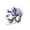
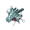
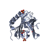
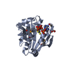
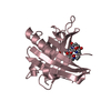
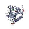
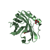
Crystal structure of Siderocalin (NGAL, Lipocalin 2) complexed with Ferric 4-methyl-catechol
Components
Neutrophil gelatinase-associated lipocalin
Keywords
TRANSPORT PROTEIN / 8-stranded anti-parallel beta barrel / 310-helix / Glycoprotein / Pyrrolidone carboxylic acid / Secreted
Function / homology
Function and homology information
positive regulation of iron ion import across plasma membrane / positive regulation of hippocampal neuron apoptotic process / positive regulation of endothelial tube morphogenesis / negative regulation of hippocampal neuron apoptotic process / positive regulation of cell projection organization / Metal sequestration by antimicrobial proteins / response to mycotoxin / response to kainic acid / siderophore transport / cellular response to increased oxygen levels ...positive regulation of iron ion import across plasma membrane / positive regulation of hippocampal neuron apoptotic process / positive regulation of endothelial tube morphogenesis / negative regulation of hippocampal neuron apoptotic process / positive regulation of cell projection organization / Metal sequestration by antimicrobial proteins / response to mycotoxin / response to kainic acid / siderophore transport / cellular response to increased oxygen levels / response to blue light / cellular response to X-ray / response to fructose / short-term memory / cellular response to interleukin-6 / enterobactin binding / iron ion sequestering activity / response to herbicide / response to iron(II) ion / positive regulation of reactive oxygen species biosynthetic process / cellular response to interleukin-1 / long-term memory / extrinsic apoptotic signaling pathway in absence of ligand / positive regulation of endothelial cell migration / cellular response to nutrient levels / acute-phase response / cellular response to nerve growth factor stimulus / Iron uptake and transport / specific granule lumen / cellular response to tumor necrosis factor / cellular response to amyloid-beta / response to virus / cellular response to hydrogen peroxide / positive regulation of cold-induced thermogenesis / cellular response to lipopolysaccharide / protease binding / Interleukin-4 and Interleukin-13 signaling / cellular response to hypoxia / defense response to bacterium / iron ion binding / response to xenobiotic stimulus / innate immune response / Neutrophil degranulation / positive regulation of gene expression / : / extracellular exosome / extracellular region / identical protein binding Similarity search - Function
Resolution: 2.3→40.62 Å / Cor.coef. Fo:Fc: 0.924 / Cor.coef. Fo:Fc free: 0.898 / SU B: 6.761 / SU ML: 0.17 / Cross valid method: THROUGHOUT / ESU R: 0.31 / ESU R Free: 0.252 / Stereochemistry target values: MAXIMUM LIKELIHOOD / Details: HYDROGENS HAVE BEEN ADDED IN THE RIDING POSITIONS
Rfactor
Num. reflection
% reflection
Selection details
Rfree
0.28973
3403
9.5 %
RANDOM
Rwork
0.24398
-
-
-
obs
0.24825
32519
99.85 %
-
Solvent computation
Ion probe radii: 0.8 Å / Shrinkage radii: 0.8 Å / VDW probe radii: 1.2 Å / Solvent model: MASK
Displacement parameters
Biso mean: 35.513 Å2
Baniso -1
Baniso -2
Baniso -3
1-
0.22 Å2
0 Å2
0 Å2
2-
-
0.22 Å2
0 Å2
3-
-
-
-0.44 Å2
Refinement step
Cycle: LAST / Resolution: 2.3→40.62 Å
Protein
Nucleic acid
Ligand
Solvent
Total
Num. atoms
3901
0
92
268
4261
Refine LS restraints
Refine-ID
Type
Dev ideal
Dev ideal target
Number
X-RAY DIFFRACTION
r_bond_refined_d
0.005
0.022
4154
X-RAY DIFFRACTION
r_bond_other_d
0.002
0.02
2813
X-RAY DIFFRACTION
r_angle_refined_deg
0.941
1.97
5641
X-RAY DIFFRACTION
r_angle_other_deg
0.791
3.002
6850
X-RAY DIFFRACTION
r_dihedral_angle_1_deg
5.172
5
528
X-RAY DIFFRACTION
r_dihedral_angle_2_deg
32.356
24.251
167
X-RAY DIFFRACTION
r_dihedral_angle_3_deg
15.069
15
629
X-RAY DIFFRACTION
r_dihedral_angle_4_deg
21.478
15
14
X-RAY DIFFRACTION
r_chiral_restr
0.061
0.2
612
X-RAY DIFFRACTION
r_gen_planes_refined
0.002
0.02
4597
X-RAY DIFFRACTION
r_gen_planes_other
0.001
0.02
857
X-RAY DIFFRACTION
r_nbd_refined
0.163
0.2
642
X-RAY DIFFRACTION
r_nbd_other
0.181
0.2
2763
X-RAY DIFFRACTION
r_nbtor_refined
0.176
0.2
1939
X-RAY DIFFRACTION
r_nbtor_other
0.08
0.2
2270
X-RAY DIFFRACTION
r_xyhbond_nbd_refined
0.094
0.2
272
X-RAY DIFFRACTION
r_metal_ion_refined
0.236
0.2
1
X-RAY DIFFRACTION
r_symmetry_vdw_refined
0.106
0.2
19
X-RAY DIFFRACTION
r_symmetry_vdw_other
0.144
0.2
47
X-RAY DIFFRACTION
r_symmetry_hbond_refined
0.113
0.2
16
X-RAY DIFFRACTION
r_mcbond_it
0.418
2
2596
X-RAY DIFFRACTION
r_mcbond_other
0.038
2
1031
X-RAY DIFFRACTION
r_mcangle_it
0.784
3
4179
X-RAY DIFFRACTION
r_scbond_it
0.758
4
1617
X-RAY DIFFRACTION
r_scangle_it
1.257
6
1450
LS refinement shell
Resolution: 2.301→2.361 Å / Total num. of bins used: 20
Rfactor
Num. reflection
% reflection
Rfree
0.349
244
-
Rwork
0.304
2320
-
obs
-
-
99.34 %
+
About Yorodumi
-
News
-
Feb 9, 2022. New format data for meta-information of EMDB entries
New format data for meta-information of EMDB entries
Version 3 of the EMDB header file is now the official format.
The previous official version 1.9 will be removed from the archive.
In the structure databanks used in Yorodumi, some data are registered as the other names, "COVID-19 virus" and "2019-nCoV". Here are the details of the virus and the list of structure data.
Jan 31, 2019. EMDB accession codes are about to change! (news from PDBe EMDB page)
EMDB accession codes are about to change! (news from PDBe EMDB page)
The allocation of 4 digits for EMDB accession codes will soon come to an end. Whilst these codes will remain in use, new EMDB accession codes will include an additional digit and will expand incrementally as the available range of codes is exhausted. The current 4-digit format prefixed with “EMD-” (i.e. EMD-XXXX) will advance to a 5-digit format (i.e. EMD-XXXXX), and so on. It is currently estimated that the 4-digit codes will be depleted around Spring 2019, at which point the 5-digit format will come into force.
The EM Navigator/Yorodumi systems omit the EMD- prefix.
Related info.:Q: What is EMD? / ID/Accession-code notation in Yorodumi/EM Navigator
Yorodumi is a browser for structure data from EMDB, PDB, SASBDB, etc.
This page is also the successor to EM Navigator detail page, and also detail information page/front-end page for Omokage search.
The word "yorodu" (or yorozu) is an old Japanese word meaning "ten thousand". "mi" (miru) is to see.
Related info.:EMDB / PDB / SASBDB / Comparison of 3 databanks / Yorodumi Search / Aug 31, 2016. New EM Navigator & Yorodumi / Yorodumi Papers / Jmol/JSmol / Function and homology information / Changes in new EM Navigator and Yorodumi
 Movie
Movie Controller

 Yorodumi
 Open data
 Basic information
 Components
 Keywords
 Function and homology information
 Homo sapiens (human)
 X-RAY DIFFRACTION /
  Authors
 Citation
 Structure visualization
 Downloads & links
 Other downloads


 PDBj
PDBj

 Assembly


 Mass: 55.845 Da / Num. of mol.: 3 / Source method: obtained synthetically / Formula: Fe
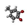
 Mass: 124.137 Da / Num. of mol.: 3 / Source method: obtained synthetically / Formula: C7H8O2
 Mass: 22.990 Da / Num. of mol.: 7 / Source method: obtained synthetically / Formula: Na
 Mass: 35.453 Da / Num. of mol.: 1 / Source method: obtained synthetically / Formula: Cl
 Mass: 92.094 Da / Num. of mol.: 9 / Source method: obtained synthetically / Formula: C3H8O3
 Sample preparation
 / Beamline: 5.0.1 / Wavelength: 1 Å
 Processing