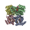
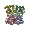
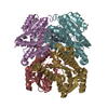
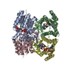
Entry Database : PDB / ID : 4z9xTitle Crystal structure of 2-keto-3-deoxy-D-gluconate dehydrogenase from Streptococcus pyogenes Gluconate 5-dehydrogenase Keywords / / / / Function / homology Function Domain/homology Component
/ / / / / / / / Biological species Streptococcus pyogenes serotype M1 (bacteria)Method / / / Resolution : 1.7 Å Authors Maruyama, Y. / Takase, R. / Oiki, S. / Mikami, B. / Murata, K. / Hashimoto, W. Funding support Organization Grant number Country Grants-in-aid from the Japan Society for the Promotion of Science Program for Promotion of Basic Research Activities for Innovative Biosciences (PROBRAIN) of Japan Targeted Proteins Research Program from the Ministry of Education, Culture, Sports, Science, and Technology (MEXT) of Japan Research fellowships from the Japan Society for the Promotion of Science for Young Scientists
Journal : Proteins / Year : 2016Title : Structural determinants in bacterial 2-keto-3-deoxy-D-gluconate dehydrogenase KduD for dual-coenzyme specificityAuthors : Takase, R. / Maruyama, Y. / Oiki, S. / Mikami, B. / Murata, K. / Hashimoto, W. History Deposition Apr 13, 2015 Deposition site / Processing site Revision 1.0 Apr 29, 2015 Provider / Type Revision 1.1 Jun 22, 2016 Group Revision 1.2 Feb 19, 2020 Group / Database references / Derived calculationsCategory / diffrn_source / pdbx_struct_oper_listItem / _diffrn_source.pdbx_synchrotron_site / _pdbx_struct_oper_list.symmetry_operationRevision 1.3 Nov 8, 2023 Group / Database references / Refinement descriptionCategory chem_comp_atom / chem_comp_bond ... chem_comp_atom / chem_comp_bond / database_2 / pdbx_initial_refinement_model Item / _database_2.pdbx_database_accession
Show all Show less
 Movie
Movie Controller

 Yorodumi
 Open data
 Basic information
 Components
 Keywords
 Function and homology information
 Streptococcus pyogenes serotype M1 (bacteria)
 X-RAY DIFFRACTION /
  Authors
 Japan, 4items
  Citation
 Structure visualization
 Downloads & links
 Other downloads


 PDBj
PDBj


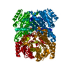
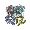
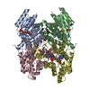
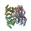
 Assembly
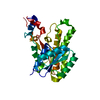
 Mass: 18.015 Da / Num. of mol.: 96 / Source method: isolated from a natural source / Formula: H2O
 Sample preparation
 Processing