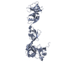
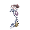
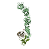
Entry Database : PDB / ID : 4z96Title Crystal structure of DNMT1 in complex with USP7 DNA (cytosine-5)-methyltransferase 1 Ubiquitin carboxyl-terminal hydrolase 7 Keywords / / Function / homology Function Domain/homology Component
/ / / / / / / / / / / / / / / / / / / / / / / / / / / / / / / / / / / / / / / / / / / / / / / / / / / / / / / / / / / / / / / / / / / / / / / / / / / / / / / / / / / / / / / / / / / / / / / / / / / / / / / / / / / / / / / / / / / / / / / / / / / / / / / / / / / / / / / Biological species Homo sapiens (human)Method / / / Resolution : 2.85 Å Authors Zhang, Z.M. / Song, J. Journal : To Be Published Title : Crystal structure of DNMT1 in complex with USP7Authors : Zhang, Z.M. / Song, J. History Deposition Apr 9, 2015 Deposition site / Processing site Revision 1.0 Oct 12, 2016 Provider / Type Revision 1.1 Sep 27, 2023 Group Data collection / Database references ... Data collection / Database references / Derived calculations / Refinement description Category chem_comp_atom / chem_comp_bond ... chem_comp_atom / chem_comp_bond / database_2 / pdbx_initial_refinement_model / pdbx_struct_oper_list Item / _database_2.pdbx_database_accession / _pdbx_struct_oper_list.symmetry_operation
Show all Show less
 Movie
Movie Controller


 Open data
 Basic information
 Components
 Keywords
 Function and homology information
 Homo sapiens (human)
 X-RAY DIFFRACTION /
  Authors
 Citation
 Structure visualization
 Downloads & links
 Other downloads
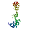


 PDBj
PDBj


 Assembly

 Sample preparation
 / Beamline: 5.0.1 / Wavelength: 0.9774 Å
 Processing