 Movie
Movie Controller

[English] 日本語
 Yorodumi
- PDB-4z5s: Crystal structure of apo-form of aldehyde deformylating oxygenase... -
+ Open data

- Basic information
| Entry | Database: PDB / ID: 4z5s | ||||||
|---|---|---|---|---|---|---|---|
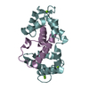
| Title | Crystal structure of apo-form of aldehyde deformylating oxygenase from Synechocystis sp.PCC 6803 | ||||||
 Components Components | Aldehyde decarbonylase | ||||||
 Keywords Keywords | LYASE / oxygenase | ||||||
| Function / homology |  Function and homology information Function and homology informationaldehyde oxygenase (deformylating) activity / aldehyde oxygenase (deformylating) / transition metal ion binding Similarity search - Function | ||||||
| Biological species |  | ||||||
| Method |  X-RAY DIFFRACTION / SYNCHROTRON / MOLECULAR REPLACEMENT / Resolution: 1.582 Å X-RAY DIFFRACTION / SYNCHROTRON / MOLECULAR REPLACEMENT / Resolution: 1.582 Å | ||||||
 Authors Authors | Jia, C. / Li, M. / Chang, W. | ||||||
 Citation Citation | Journal: BMC Biotechnol. / Year: 2017 Title: Identification of residues important for the activity of aldehyde-deformylating oxygenase through investigation into the structure-activity relationship Authors: Wang, Q. / Bao, L. / Jia, C. / Li, M. / Li, J.J. / Lu, X. | ||||||
| History |
|
- Structure visualization
| Structure viewer | Molecule: MolmilJmol/JSmol |
|---|
- Downloads & links
-Download
| PDBx/mmCIF format | 4z5s.cif.gz | 115.7 KB | Display | PDBx/mmCIF format |
|---|---|---|---|---|
| PDB format | pdb4z5s.ent.gz | 89.1 KB | Display | PDB format |
| PDBx/mmJSON format | 4z5s.json.gz | Tree view | PDBx/mmJSON format | |
| Others |  Other downloads Other downloads |
-Validation report
| Summary document | 4z5s_validation.pdf.gz | 419.1 KB | Display | wwPDB validaton report |
|---|---|---|---|---|
| Full document | 4z5s_full_validation.pdf.gz | 419 KB | Display | |
| Data in XML | 4z5s_validation.xml.gz | 12.6 KB | Display | |
| Data in CIF | 4z5s_validation.cif.gz | 19 KB | Display | |
| Arichive directory | https://data.pdbj.org/pub/pdb/validation_reports/z5/4z5sftp://data.pdbj.org/pub/pdb/validation_reports/z5/4z5s | HTTPS FTP |
-Related structure data
| Related structure data |  2oc5S S: Starting model for refinement |
|---|---|
| Similar structure data |


-Links
 PDBj
PDBj


- Assembly
| Deposited unit | 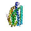
| ||||||||
|---|---|---|---|---|---|---|---|---|---|
| 1 |
| ||||||||
| Unit cell |
|
-Components
| #1: Protein | Mass: 26202.646 Da / Num. of mol.: 1 Source method: isolated from a genetically manipulated source Source: (gene. exp.) Strain: PCC 6803 / Kazusa / Gene: sll0208 / Production host: References: UniProt: Q55688, aldehyde oxygenase (deformylating) |
|---|---|
| #2: Water | ChemComp-HOH /  Mass: 18.015 Da / Num. of mol.: 282 / Source method: isolated from a natural source / Formula: H2O Mass: 18.015 Da / Num. of mol.: 282 / Source method: isolated from a natural source / Formula: H2O |
-Experimental details
-Experiment
| Experiment | Method: X-RAY DIFFRACTION |
|---|
- Sample preparation
| Crystal | Density Matthews: 2.09 Å3/Da / Density % sol: 41.04 % Description: THE ENTRY CONTAINS FRIEDEL PAIRS IN F_PLUS/MINUS COLUMNS. |
|---|---|
| Crystal grow | Temperature: 291 K / Method: vapor diffusion, sitting drop / pH: 5.2 Details: 19%(v/v) 2-Propanol, 0.2M Ammonium sulfate, 0.1M Sodium citrate tribasic dehydrate pH5.2, 18% PEG3350 |
-Data collection
| Diffraction | Mean temperature: 100 K |
|---|---|
| Diffraction source | Source: SYNCHROTRON / Site: SSRF  / Beamline: BL17U / Wavelength: 0.98 Å / Beamline: BL17U / Wavelength: 0.98 Å |
| Detector | Type: ADSC QUANTUM 315 / Detector: CCD / Date: Jan 8, 2012 |
| Radiation | Monochromator: GRAPHITE / Protocol: SINGLE WAVELENGTH / Monochromatic (M) / Laue (L): M / Scattering type: x-ray |
| Radiation wavelength | Wavelength: 0.98 Å / Relative weight: 1 |
| Reflection | Resolution: 1.582→44.014 Å / Num. obs: 29825 / % possible obs: 99.6 % / Redundancy: 8.5 % / Net I/σ(I): 22.2 |
| Reflection shell | Resolution: 1.58→1.61 Å / Redundancy: 9.1 % / Rmerge(I) obs: 0.497 / Mean I/σ(I) obs: 4.75 / % possible all: 99.7 |
- Processing
| Software |
| ||||||||||||||||||||||||||||||||||||||||||||||||||||||||||||||||||||||||||||||||||||
|---|---|---|---|---|---|---|---|---|---|---|---|---|---|---|---|---|---|---|---|---|---|---|---|---|---|---|---|---|---|---|---|---|---|---|---|---|---|---|---|---|---|---|---|---|---|---|---|---|---|---|---|---|---|---|---|---|---|---|---|---|---|---|---|---|---|---|---|---|---|---|---|---|---|---|---|---|---|---|---|---|---|---|---|---|---|
| Refinement | Method to determine structure: MOLECULAR REPLACEMENT Starting model: 2OC5 Resolution: 1.582→44.014 Å / SU ML: 0.13 / Cross valid method: THROUGHOUT / σ(F): 1.36 / Phase error: 19.55 / Stereochemistry target values: ML Details: SF FILE CONTAINS FRIEDEL PAIRS UNDER I/F_MINUS AND I/F_PLUS COLUMNS.
| ||||||||||||||||||||||||||||||||||||||||||||||||||||||||||||||||||||||||||||||||||||
| Solvent computation | Shrinkage radii: 0.9 Å / VDW probe radii: 1.11 Å / Solvent model: FLAT BULK SOLVENT MODEL | ||||||||||||||||||||||||||||||||||||||||||||||||||||||||||||||||||||||||||||||||||||
| Refinement step | Cycle: LAST / Resolution: 1.582→44.014 Å
| ||||||||||||||||||||||||||||||||||||||||||||||||||||||||||||||||||||||||||||||||||||
| Refine LS restraints |
| ||||||||||||||||||||||||||||||||||||||||||||||||||||||||||||||||||||||||||||||||||||
| LS refinement shell |
| ||||||||||||||||||||||||||||||||||||||||||||||||||||||||||||||||||||||||||||||||||||
| Refinement TLS params. | Method: refined / Origin x: 14.2667 Å / Origin y: 102.1311 Å / Origin z: 46.6198 Å
| ||||||||||||||||||||||||||||||||||||||||||||||||||||||||||||||||||||||||||||||||||||
| Refinement TLS group | Selection details: all |
