 Movie
Movie Controller


+ Open data

- Basic information
| Entry | Database: PDB / ID: 4yxq | ||||||
|---|---|---|---|---|---|---|---|
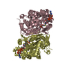
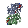
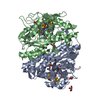
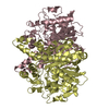
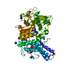
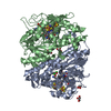
| Title | PksG, a HMG-CoA Synthase from Bacillus subtilis | ||||||
 Components Components | Polyketide biosynthesis 3-hydroxy-3-methylglutaryl-ACP synthase PksG | ||||||
 Keywords Keywords | TRANSFERASE / polyketide / bacillaene | ||||||
| Function / homology |  Function and homology information Function and homology informationTransferases; Acyltransferases; Acyl groups converted into alkyl groups on transfer / farnesyl diphosphate biosynthetic process, mevalonate pathway / hydroxymethylglutaryl-CoA synthase activity / acetyl-CoA metabolic process / antibiotic biosynthetic process / cytoplasm Similarity search - Function | ||||||
| Biological species |  | ||||||
| Method |  X-RAY DIFFRACTION / SYNCHROTRON / Resolution: 2.47 Å X-RAY DIFFRACTION / SYNCHROTRON / Resolution: 2.47 Å | ||||||
 Authors Authors | Nair, A.V. / Race, P.R. / Till, M. | ||||||
 Citation Citation | Journal: To Be Published Title: PksG, a HMG-CoA Synthase from Bacillus subtilis Authors: Nair, A.V. / Race, P.R. / Till, M. | ||||||
| History |
|
- Structure visualization
| Structure viewer | Molecule: MolmilJmol/JSmol |
|---|
- Downloads & links
-Download
| PDBx/mmCIF format | 4yxq.cif.gz | 322.1 KB | Display | PDBx/mmCIF format |
|---|---|---|---|---|
| PDB format | pdb4yxq.ent.gz | 261.4 KB | Display | PDB format |
| PDBx/mmJSON format | 4yxq.json.gz | Tree view | PDBx/mmJSON format | |
| Others |  Other downloads Other downloads |
-Validation report
| Arichive directory | https://data.pdbj.org/pub/pdb/validation_reports/yx/4yxqftp://data.pdbj.org/pub/pdb/validation_reports/yx/4yxq | HTTPS FTP |
|---|
-Related structure data
| Similar structure data |
|---|


-Links
 PDBj
PDBj

- Assembly
| Deposited unit | 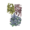
| ||||||||||||||||||||||||||||||||||||||||||||||||||||||||||||||||||||||||||||||||||||||||||||||||||
|---|---|---|---|---|---|---|---|---|---|---|---|---|---|---|---|---|---|---|---|---|---|---|---|---|---|---|---|---|---|---|---|---|---|---|---|---|---|---|---|---|---|---|---|---|---|---|---|---|---|---|---|---|---|---|---|---|---|---|---|---|---|---|---|---|---|---|---|---|---|---|---|---|---|---|---|---|---|---|---|---|---|---|---|---|---|---|---|---|---|---|---|---|---|---|---|---|---|---|---|
| 1 | 
| ||||||||||||||||||||||||||||||||||||||||||||||||||||||||||||||||||||||||||||||||||||||||||||||||||
| 2 | 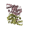
| ||||||||||||||||||||||||||||||||||||||||||||||||||||||||||||||||||||||||||||||||||||||||||||||||||
| Unit cell |
| ||||||||||||||||||||||||||||||||||||||||||||||||||||||||||||||||||||||||||||||||||||||||||||||||||
| Noncrystallographic symmetry (NCS) | NCS domain:
NCS domain segments: Component-ID: _ / Beg auth comp-ID: VAL / Beg label comp-ID: VAL / End auth comp-ID: SER / End label comp-ID: SER / Refine code: _ / Auth seq-ID: 2 - 420 / Label seq-ID: 2 - 420
NCS ensembles :
|
-Components
| #1: Protein | Mass: 46829.164 Da / Num. of mol.: 4 Source method: isolated from a genetically manipulated source Source: (gene. exp.) Strain: 168 / Gene: pksG, BSU17150 / Production host: References: UniProt: P40830, Transferases; Acyltransferases; Acyl groups converted into alkyl groups on transfer #2: Chemical |   Mass: 106.120 Da / Num. of mol.: 3 / Source method: obtained synthetically / Formula: C4H10O3 Mass: 106.120 Da / Num. of mol.: 3 / Source method: obtained synthetically / Formula: C4H10O3#3: Water | ChemComp-HOH / |  Mass: 18.015 Da / Num. of mol.: 357 / Source method: isolated from a natural source / Formula: H2O Mass: 18.015 Da / Num. of mol.: 357 / Source method: isolated from a natural source / Formula: H2O |
|---|
-Experimental details
-Experiment
| Experiment | Method: X-RAY DIFFRACTION / Number of used crystals: 1 |
|---|
- Sample preparation
| Crystal | Density Matthews: 2.41 Å3/Da / Density % sol: 48.97 % |
|---|---|
| Crystal grow | Temperature: 293 K / Method: vapor diffusion, hanging drop / pH: 9 / Details: 0.1 M MMT, 25% PEG 1500, 30% glycerol |
-Data collection
| Diffraction | Mean temperature: 100 K | |||||||||||||||||||||||||||
|---|---|---|---|---|---|---|---|---|---|---|---|---|---|---|---|---|---|---|---|---|---|---|---|---|---|---|---|---|
| Diffraction source | Source: SYNCHROTRON / Site: Diamond  / Beamline: I04 / Wavelength: 0.9795 Å / Beamline: I04 / Wavelength: 0.9795 Å | |||||||||||||||||||||||||||
| Detector | Type: ADSC QUANTUM 315 / Detector: CCD / Date: Feb 27, 2012 | |||||||||||||||||||||||||||
| Radiation | Protocol: SINGLE WAVELENGTH / Monochromatic (M) / Laue (L): M / Scattering type: x-ray | |||||||||||||||||||||||||||
| Radiation wavelength | Wavelength: 0.9795 Å / Relative weight: 1 | |||||||||||||||||||||||||||
| Reflection | Resolution: 2.47→69.84 Å / Num. obs: 65451 / % possible obs: 100 % / Redundancy: 7 % / CC1/2: 0.985 / Rmerge(I) obs: 0.253 / Rpim(I) all: 0.103 / Net I/σ(I): 6.2 / Num. measured all: 458826 / Scaling rejects: 1685 | |||||||||||||||||||||||||||
| Reflection shell | Diffraction-ID: 1 / Rejects: _
|
- Processing
| Software |
| |||||||||||||||||||||||||||||||||||||||||||||||||||||||||||||||||
|---|---|---|---|---|---|---|---|---|---|---|---|---|---|---|---|---|---|---|---|---|---|---|---|---|---|---|---|---|---|---|---|---|---|---|---|---|---|---|---|---|---|---|---|---|---|---|---|---|---|---|---|---|---|---|---|---|---|---|---|---|---|---|---|---|---|---|
| Refinement | Resolution: 2.47→63.74 Å / Cor.coef. Fo:Fc: 0.903 / Cor.coef. Fo:Fc free: 0.861 / WRfactor Rfree: 0.2526 / WRfactor Rwork: 0.2096 / FOM work R set: 0.797 / SU B: 10.014 / SU ML: 0.223 / SU R Cruickshank DPI: 0.6604 / SU Rfree: 0.3124 / Cross valid method: THROUGHOUT / σ(F): 0 / ESU R: 0.642 / ESU R Free: 0.312 / Stereochemistry target values: MAXIMUM LIKELIHOOD Details: HYDROGENS HAVE BEEN ADDED IN THE RIDING POSITIONS U VALUES : REFINED INDIVIDUALLY
| |||||||||||||||||||||||||||||||||||||||||||||||||||||||||||||||||
| Solvent computation | Ion probe radii: 0.8 Å / Shrinkage radii: 0.8 Å / VDW probe radii: 1.2 Å / Solvent model: MASK | |||||||||||||||||||||||||||||||||||||||||||||||||||||||||||||||||
| Displacement parameters | Biso max: 120.25 Å2 / Biso mean: 32.254 Å2 / Biso min: 5.62 Å2
| |||||||||||||||||||||||||||||||||||||||||||||||||||||||||||||||||
| Refinement step | Cycle: final / Resolution: 2.47→63.74 Å
| |||||||||||||||||||||||||||||||||||||||||||||||||||||||||||||||||
| Refine LS restraints |
| |||||||||||||||||||||||||||||||||||||||||||||||||||||||||||||||||
| Refine LS restraints NCS | Refine-ID: X-RAY DIFFRACTION / Type: interatomic distance / Weight position: 0.05
| |||||||||||||||||||||||||||||||||||||||||||||||||||||||||||||||||
| LS refinement shell | Resolution: 2.475→2.539 Å / Total num. of bins used: 20
|
