 Movie
Movie Controller

[English] 日本語
 Yorodumi
- PDB-1s5o: Structural and Mutational Characterization of L-carnitine Binding... -
+ Open data

- Basic information
| Entry | Database: PDB / ID: 1s5o | ||||||
|---|---|---|---|---|---|---|---|
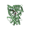
| Title | Structural and Mutational Characterization of L-carnitine Binding to Human carnitine Acetyltransferase | ||||||
 Components Components | carnitine acetyltransferase isoform 2 | ||||||
 Keywords Keywords | TRANSFERASE / Carnitine acetyltransferase / binary complex / steady-state enzyme kinetics / substrate binding site | ||||||
| Function / homology |  Function and homology information Function and homology informationcarnitine O-acetyltransferase / carnitine O-acetyltransferase activity / Beta-oxidation of pristanoyl-CoA / carnitine O-octanoyltransferase / acyl-CoA oxidase activity / carnitine O-octanoyltransferase activity / carnitine metabolic process, CoA-linked / short-chain fatty acid metabolic process / medium-chain fatty acid metabolic process / fatty acid beta-oxidation using acyl-CoA oxidase ...carnitine O-acetyltransferase / carnitine O-acetyltransferase activity / Beta-oxidation of pristanoyl-CoA / carnitine O-octanoyltransferase / acyl-CoA oxidase activity / carnitine O-octanoyltransferase activity / carnitine metabolic process, CoA-linked / short-chain fatty acid metabolic process / medium-chain fatty acid metabolic process / fatty acid beta-oxidation using acyl-CoA oxidase / peroxisomal matrix / Peroxisomal protein import / peroxisome / mitochondrial inner membrane / endoplasmic reticulum / mitochondrion / cytosol Similarity search - Function | ||||||
| Biological species |  Homo sapiens (human) Homo sapiens (human) | ||||||
| Method |  X-RAY DIFFRACTION / SYNCHROTRON / MOLECULAR REPLACEMENT / Resolution: 1.8 Å X-RAY DIFFRACTION / SYNCHROTRON / MOLECULAR REPLACEMENT / Resolution: 1.8 Å | ||||||
 Authors Authors | Govindasamy, L. / Kukar, T. / Lian, W. / Pedersen, B. / Gu, Y. / Agbandje-Mckenna, M. / Jin, S. / Mckenna, R. / Wu, D. | ||||||
 Citation Citation | Journal: J.Struct.Biol. / Year: 2004 Title: Structural and mutational characterization of l-carnitine binding to human carnitine acetyltransferase. Authors: Govindasamy, L. / Kukar, T. / Lian, W. / Pedersen, B. / Gu, Y. / Agbandje-McKenna, M. / Jin, S. / McKenna, R. / Wu, D. | ||||||
| History |
| ||||||
| Remark 999 | SEQUENCE THE AUTHORS STATE THE ELECTRON DENSITY MAP CONFIRMS THE THR RESIDUE. |
- Structure visualization
| Structure viewer | Molecule: MolmilJmol/JSmol |
|---|
- Downloads & links
-Download
| PDBx/mmCIF format | 1s5o.cif.gz | 140.5 KB | Display | PDBx/mmCIF format |
|---|---|---|---|---|
| PDB format | pdb1s5o.ent.gz | 108.4 KB | Display | PDB format |
| PDBx/mmJSON format | 1s5o.json.gz | Tree view | PDBx/mmJSON format | |
| Others |  Other downloads Other downloads |
-Validation report
| Arichive directory | https://data.pdbj.org/pub/pdb/validation_reports/s5/1s5oftp://data.pdbj.org/pub/pdb/validation_reports/s5/1s5o | HTTPS FTP |
|---|
-Related structure data
| Related structure data |  1nm8S S: Starting model for refinement |
|---|---|
| Similar structure data |

-Links
 PDBj
PDBj

- Assembly
| Deposited unit | 
| ||||||||
|---|---|---|---|---|---|---|---|---|---|
| 1 |
| ||||||||
| Unit cell |
|
-Components
| #1: Protein | Mass: 69902.867 Da / Num. of mol.: 1 Source method: isolated from a genetically manipulated source Source: (gene. exp.) Homo sapiens (human) / Production host:  |
|---|---|
| #2: Chemical | ChemComp-152 /   Mass: 162.207 Da / Num. of mol.: 1 / Source method: obtained synthetically / Formula: C7H16NO3 Mass: 162.207 Da / Num. of mol.: 1 / Source method: obtained synthetically / Formula: C7H16NO3 |
| #3: Water | ChemComp-HOH /  Mass: 18.015 Da / Num. of mol.: 465 / Source method: isolated from a natural source / Formula: H2O Mass: 18.015 Da / Num. of mol.: 465 / Source method: isolated from a natural source / Formula: H2O |
-Experimental details
-Experiment
| Experiment | Method: X-RAY DIFFRACTION / Number of used crystals: 1 |
|---|
- Sample preparation
| Crystal | Density Matthews: 2.39 Å3/Da / Density % sol: 48.49 % | ||||||||||||||||||||||||||||||||||||||||||
|---|---|---|---|---|---|---|---|---|---|---|---|---|---|---|---|---|---|---|---|---|---|---|---|---|---|---|---|---|---|---|---|---|---|---|---|---|---|---|---|---|---|---|---|
| Crystal grow | Temperature: 295 K / Method: vapor diffusion, hanging drop / pH: 6.2 Details: Bis-Tris, NaCl, PEG8000, L-Carnitine, hpCAT, pH 6.2, VAPOR DIFFUSION, HANGING DROP, temperature 295K | ||||||||||||||||||||||||||||||||||||||||||
| Crystal grow | *PLUS Method: vapor diffusion | ||||||||||||||||||||||||||||||||||||||||||
| Components of the solutions | *PLUS
|
-Data collection
| Diffraction | Mean temperature: 100 K |
|---|---|
| Diffraction source | Source: SYNCHROTRON / Site: CHESS  / Beamline: F1 / Wavelength: 0.9504 Å / Beamline: F1 / Wavelength: 0.9504 Å |
| Detector | Type: ADSC QUANTUM 4 / Detector: CCD |
| Radiation | Protocol: SINGLE WAVELENGTH / Monochromatic (M) / Laue (L): M / Scattering type: x-ray |
| Radiation wavelength | Wavelength: 0.9504 Å / Relative weight: 1 |
| Reflection | Resolution: 1.8→50 Å / Num. obs: 58948 / % possible obs: 91.6 % / Observed criterion σ(F): 4 / Rsym value: 0.086 / Net I/σ(I): 4.12 |
| Reflection shell | Resolution: 1.8→1.86 Å / Num. unique all: 5280 / Rsym value: 0.343 / % possible all: 83.3 |
| Reflection | *PLUS Lowest resolution: 50 Å / Rmerge(I) obs: 0.086 |
| Reflection shell | *PLUS Highest resolution: 1.8 Å / % possible obs: 83.3 % / Rmerge(I) obs: 0.343 |
- Processing
| Software |
| ||||||||||||
|---|---|---|---|---|---|---|---|---|---|---|---|---|---|
| Refinement | Method to determine structure: MOLECULAR REPLACEMENT Starting model: 1NM8 Resolution: 1.8→50 Å / σ(F): 4 / Stereochemistry target values: Engh & Huber
| ||||||||||||
| Refinement step | Cycle: LAST / Resolution: 1.8→50 Å
| ||||||||||||
| Refine LS restraints |
| ||||||||||||
| Software | *PLUS Name: SHELXL / Version: 97 / Classification: refinement | ||||||||||||
| Refinement | *PLUS Lowest resolution: 50 Å / σ(F): 5 / Rfactor Rfree: 0.223 | ||||||||||||
| Solvent computation | *PLUS | ||||||||||||
| Displacement parameters | *PLUS | ||||||||||||
| Refine LS restraints | *PLUS
|
