 Movie
Movie Controller

[English] 日本語
 Yorodumi
- PDB-4yrq: Crystal structure of T. cruzi Histidyl-tRNA synthetase in complex... -
+ Open data

- Basic information
| Entry | Database: PDB / ID: 4yrq | ||||||
|---|---|---|---|---|---|---|---|
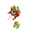
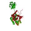
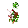
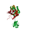
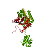
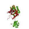
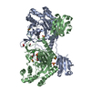
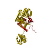
| Title | Crystal structure of T. cruzi Histidyl-tRNA synthetase in complex with 6-amino-2H-chromen-2-one (Chem 744) | ||||||
 Components Components | Histidyl-tRNA synthetase | ||||||
 Keywords Keywords | Ligase/Ligase inhibitor / ligase / aminoacyl-tRNA synthetase / aaRS / HisRS / Trypanosoma cruzi / protein-inhibitor complex / Ligase-Ligase inhibitor complex | ||||||
| Function / homology |  Function and homology information Function and homology informationhistidine-tRNA ligase / histidine-tRNA ligase activity / histidyl-tRNA aminoacylation / ATP binding / cytoplasm Similarity search - Function | ||||||
| Biological species |  | ||||||
| Method |  X-RAY DIFFRACTION / MOLECULAR REPLACEMENT / Resolution: 2.05 Å X-RAY DIFFRACTION / MOLECULAR REPLACEMENT / Resolution: 2.05 Å | ||||||
 Authors Authors | Koh, C.-Y. / Hol, W.G.J. | ||||||
 Citation Citation | Journal: Acta Crystallogr. D Biol. Crystallogr. / Year: 2015 Title: A binding hotspot in Trypanosoma cruzi histidyl-tRNA synthetase revealed by fragment-based crystallographic cocktail screens. Authors: Koh, C.Y. / Siddaramaiah, L.K. / Ranade, R.M. / Nguyen, J. / Jian, T. / Zhang, Z. / Gillespie, J.R. / Buckner, F.S. / Verlinde, C.L. / Fan, E. / Hol, W.G. | ||||||
| History |
|
- Structure visualization
| Structure viewer | Molecule: MolmilJmol/JSmol |
|---|
- Downloads & links
-Download
| PDBx/mmCIF format | 4yrq.cif.gz | 180.8 KB | Display | PDBx/mmCIF format |
|---|---|---|---|---|
| PDB format | pdb4yrq.ent.gz | 139.5 KB | Display | PDB format |
| PDBx/mmJSON format | 4yrq.json.gz | Tree view | PDBx/mmJSON format | |
| Others |  Other downloads Other downloads |
-Validation report
| Arichive directory | https://data.pdbj.org/pub/pdb/validation_reports/yr/4yrqftp://data.pdbj.org/pub/pdb/validation_reports/yr/4yrq | HTTPS FTP |
|---|
-Related structure data
| Related structure data |  4yp0C  4ypfC  4yrcC  4yreC  4yrfC  4yrgC  4yriC  4yrjC  4yrkC  4yrlC  4yrmC  4yrnC  4yroC  4yrpC  4yrrC  4yrsC 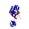 4yrtC  3lc0S C: citing same article ( S: Starting model for refinement |
|---|---|
| Similar structure data |


-Links
 PDBj
PDBj

- Assembly
| Deposited unit | 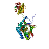
| |||||||||||||||
|---|---|---|---|---|---|---|---|---|---|---|---|---|---|---|---|---|
| 1 | 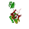
| |||||||||||||||
| Unit cell |
| |||||||||||||||
| Components on special symmetry positions |
|
-Components
-Protein , 1 types, 1 molecules A
| #1: Protein | Mass: 51185.590 Da / Num. of mol.: 1 / Fragment: UNP residues 45-478 Source method: isolated from a genetically manipulated source Source: (gene. exp.) Strain: CL Brener / Gene: Tc00.1047053507019.40 / Plasmid: AVA0421 / Production host:  |
|---|
-Non-polymers , 6 types, 209 molecules 


| #2: Chemical | ChemComp-HIS /  Type: L-peptide linking / Mass: 156.162 Da / Num. of mol.: 1 / Source method: obtained synthetically / Formula: C6H10N3O2 Type: L-peptide linking / Mass: 156.162 Da / Num. of mol.: 1 / Source method: obtained synthetically / Formula: C6H10N3O2 | ||||
|---|---|---|---|---|---|
| #3: Chemical | ChemComp-4JQ /  Mass: 161.157 Da / Num. of mol.: 1 / Source method: obtained synthetically / Formula: C9H7NO2 Mass: 161.157 Da / Num. of mol.: 1 / Source method: obtained synthetically / Formula: C9H7NO2 | ||||
| #4: Chemical | ChemComp-EDO /  Mass: 62.068 Da / Num. of mol.: 1 / Source method: obtained synthetically / Formula: C2H6O2 Mass: 62.068 Da / Num. of mol.: 1 / Source method: obtained synthetically / Formula: C2H6O2 | ||||
| #5: Chemical |  Mass: 78.133 Da / Num. of mol.: 2 / Source method: obtained synthetically / Formula: C2H6OS / Comment: DMSO, precipitant*YM Mass: 78.133 Da / Num. of mol.: 2 / Source method: obtained synthetically / Formula: C2H6OS / Comment: DMSO, precipitant*YM#6: Chemical | ChemComp-SO4 / |  Mass: 96.063 Da / Num. of mol.: 1 / Source method: obtained synthetically / Formula: SO4 Mass: 96.063 Da / Num. of mol.: 1 / Source method: obtained synthetically / Formula: SO4#7: Water | ChemComp-HOH / | Mass: 18.015 Da / Num. of mol.: 203 / Source method: isolated from a natural source / Formula: H2O |
-Experimental details
-Experiment
| Experiment | Method: X-RAY DIFFRACTION / Number of used crystals: 1 |
|---|
- Sample preparation
| Crystal | Density Matthews: 2.51 Å3/Da / Density % sol: 50.9 % |
|---|---|
| Crystal grow | Temperature: 298 K / Method: vapor diffusion, sitting drop Details: 0.2 M ammonium sulfate, 23 % to 28 % PEG 3350, 0.1 M sodium citrate pH 4.8 to 5.3, 1 mM TCEP PH range: 4.8 to 5.3 |
-Data collection
| Diffraction | Mean temperature: 100 K | |||||||||||||||||||||||||||
|---|---|---|---|---|---|---|---|---|---|---|---|---|---|---|---|---|---|---|---|---|---|---|---|---|---|---|---|---|
| Diffraction source | Source: ROTATING ANODE / Type: RIGAKU MICROMAX-007 / Wavelength: 1.54 Å | |||||||||||||||||||||||||||
| Detector | Type: RIGAKU SATURN 944 / Detector: CCD / Date: May 20, 2013 | |||||||||||||||||||||||||||
| Radiation | Monochromator: VariMax HF (Osmic) / Protocol: SINGLE WAVELENGTH / Monochromatic (M) / Laue (L): M / Scattering type: x-ray | |||||||||||||||||||||||||||
| Radiation wavelength | Wavelength: 1.54 Å / Relative weight: 1 | |||||||||||||||||||||||||||
| Reflection | Resolution: 2.05→27.82 Å / Num. obs: 31011 / % possible obs: 98.2 % / Redundancy: 2.8 % / CC1/2: 0.998 / Rmerge(I) obs: 0.055 / Rpim(I) all: 0.037 / Net I/σ(I): 10.6 / Num. measured all: 87563 | |||||||||||||||||||||||||||
| Reflection shell | Diffraction-ID: 1 / Rejects: _
|
- Processing
| Software |
| ||||||||||||||||||||||||||||||||||||||||||||||||||||||||||||||||||||||||||||||||||||||||||||||||||||||||||||||||||||||||||||||||||||||||||||||||||||||
|---|---|---|---|---|---|---|---|---|---|---|---|---|---|---|---|---|---|---|---|---|---|---|---|---|---|---|---|---|---|---|---|---|---|---|---|---|---|---|---|---|---|---|---|---|---|---|---|---|---|---|---|---|---|---|---|---|---|---|---|---|---|---|---|---|---|---|---|---|---|---|---|---|---|---|---|---|---|---|---|---|---|---|---|---|---|---|---|---|---|---|---|---|---|---|---|---|---|---|---|---|---|---|---|---|---|---|---|---|---|---|---|---|---|---|---|---|---|---|---|---|---|---|---|---|---|---|---|---|---|---|---|---|---|---|---|---|---|---|---|---|---|---|---|---|---|---|---|---|---|---|---|
| Refinement | Method to determine structure: MOLECULAR REPLACEMENT Starting model: 3LC0 Resolution: 2.05→27.82 Å / Cor.coef. Fo:Fc: 0.958 / Cor.coef. Fo:Fc free: 0.941 / SU B: 7.604 / SU ML: 0.109 / Cross valid method: THROUGHOUT / σ(F): 0 / ESU R: 0.18 / ESU R Free: 0.156 / Stereochemistry target values: MAXIMUM LIKELIHOOD Details: HYDROGENS HAVE BEEN ADDED IN THE RIDING POSITIONS U VALUES : WITH TLS ADDED
| ||||||||||||||||||||||||||||||||||||||||||||||||||||||||||||||||||||||||||||||||||||||||||||||||||||||||||||||||||||||||||||||||||||||||||||||||||||||
| Solvent computation | Ion probe radii: 0.8 Å / Shrinkage radii: 0.8 Å / VDW probe radii: 1.2 Å / Solvent model: MASK | ||||||||||||||||||||||||||||||||||||||||||||||||||||||||||||||||||||||||||||||||||||||||||||||||||||||||||||||||||||||||||||||||||||||||||||||||||||||
| Displacement parameters | Biso max: 105.36 Å2 / Biso mean: 39.798 Å2 / Biso min: 18.4 Å2
| ||||||||||||||||||||||||||||||||||||||||||||||||||||||||||||||||||||||||||||||||||||||||||||||||||||||||||||||||||||||||||||||||||||||||||||||||||||||
| Refinement step | Cycle: final / Resolution: 2.05→27.82 Å
| ||||||||||||||||||||||||||||||||||||||||||||||||||||||||||||||||||||||||||||||||||||||||||||||||||||||||||||||||||||||||||||||||||||||||||||||||||||||
| Refine LS restraints |
| ||||||||||||||||||||||||||||||||||||||||||||||||||||||||||||||||||||||||||||||||||||||||||||||||||||||||||||||||||||||||||||||||||||||||||||||||||||||
| LS refinement shell | Resolution: 2.05→2.103 Å / Total num. of bins used: 20
| ||||||||||||||||||||||||||||||||||||||||||||||||||||||||||||||||||||||||||||||||||||||||||||||||||||||||||||||||||||||||||||||||||||||||||||||||||||||
| Refinement TLS params. | Method: refined / Refine-ID: X-RAY DIFFRACTION
| ||||||||||||||||||||||||||||||||||||||||||||||||||||||||||||||||||||||||||||||||||||||||||||||||||||||||||||||||||||||||||||||||||||||||||||||||||||||
| Refinement TLS group |
|
