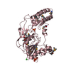
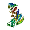
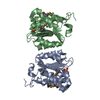
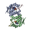
- PDB-4yod: Crystal structure of a thioredoxin-like protein (BACCAC_02376) fr... -
+
Open data
ID or keywords:
Loading...
-
Basic information
Entry
Database: PDB / ID: 4yod
Title
Crystal structure of a thioredoxin-like protein (BACCAC_02376) from Bacteroides caccae ATCC 43185 at 1.90 A resolution
Components
thioredoxin-like protein
Keywords
OXIDOREDUCTASE / Thioredoxin fold / Structural Genomics / Joint Center for Structural Genomics / JCSG / Protein Structure Initiative / PSI-BIOLOGY
Function / homology
Domain of unknown function DUF5106 / Domain of unknown function (DUF5106) / Thioredoxin-like / Thioredoxin-like fold / Thioredoxin-like superfamily / Uncharacterized protein
Function and homology information
Biological species
Bacteroides caccae ATCC 43185 (bacteria)
Method
X-RAY DIFFRACTION / SYNCHROTRON / MAD / Resolution: 1.9 Å
Mass: 18.015 Da / Num. of mol.: 197 / Source method: isolated from a natural source / Formula: H2O
Has protein modification
Y
Sequence details
THE CONSTRUCT (27-312) WAS EXPRESSED WITH A PURIFICATION TAG MGSDKIHHHHHHENLYFQG. THE TAG WAS ...THE CONSTRUCT (27-312) WAS EXPRESSED WITH A PURIFICATION TAG MGSDKIHHHHHHENLYFQG. THE TAG WAS REMOVED WITH TEV PROTEASE LEAVING ONLY A GLYCINE (0) FOLLOWED BY THE TARGET SEQUENCE.
-
Experimental details
-
Experiment
Experiment
Method: X-RAY DIFFRACTION / Number of used crystals: 1
-
Sample preparation
Crystal
Density Matthews: 2.2 Å3/Da / Density % sol: 44.06 %
Resolution: 1.9→28.343 Å / Num. obs: 22102 / % possible obs: 91.7 % / Observed criterion σ(I): -3 / Redundancy: 1.77 % / Biso Wilson estimate: 34.007 Å2 / Rmerge F obs: 0.998 / Rmerge(I) obs: 0.04 / Rrim(I) all: 0.056 / Net I/σ(I): 10.6 / Num. measured all: 72712
Reflection shell
Resolution (Å)
Highest resolution (Å)
Rmerge F obs
Rmerge(I) obs
Mean I/σ(I) obs
Num. measured obs
Num. possible
Num. unique obs
Rrim(I) all
Diffraction-ID
% possible all
1.91-1.98
0.783
0.505
2
6856
4556
4056
0.702
1
89
1.98-2.06
0.825
0.336
3
7764
4508
4244
0.467
94.1
2.06-2.15
0.891
0.238
4.5
7435
4315
4091
0.331
94.8
2.15-2.26
0.934
0.177
5.8
7230
4324
4062
0.245
93.9
2.26-2.41
0.961
0.132
7.4
7552
4825
4416
0.182
91.5
2.41-2.59
0.981
0.091
9.3
7453
4288
4053
0.126
94.5
2.59-2.85
0.991
0.062
12.2
7532
4492
4218
0.086
93.9
2.85-3.26
0.995
0.041
16.4
6841
4472
3991
0.057
89.2
3.26-4.1
0.996
0.03
21.9
7026
4472
3942
0.041
88.1
4.1
0.998
0.023
24.8
7023
4550
4029
0.032
88.5
-
Phasing
Phasing
Method: MAD
-
Processing
Software
Name
Version
Classification
PDB_EXTRACT
3.1
dataextraction
SHELX
phasing
SHARP
phasing
XSCALE
November3, 2014BUILT=20141118
datascaling
XDS
datascaling
BUSTER
2.10.2
refinement
SHELXD
phasing
Refinement
Method to determine structure: MAD / Resolution: 1.9→28.343 Å / Cor.coef. Fo:Fc: 0.9583 / Cor.coef. Fo:Fc free: 0.9395 / Occupancy max: 1 / Occupancy min: 0.5 / Cross valid method: THROUGHOUT / σ(F): 0 Details: 1. A MET-INHIBITION PROTOCOL WAS USED FOR SELENOMETHIONINE INCORPORATION DURING PROTEIN EXPRESSION. THE OCCUPANCY OF THE SE ATOMS IN THE MSE RESIDUES WAS REDUCED TO 0.75 FOR THE REDUCED ...Details: 1. A MET-INHIBITION PROTOCOL WAS USED FOR SELENOMETHIONINE INCORPORATION DURING PROTEIN EXPRESSION. THE OCCUPANCY OF THE SE ATOMS IN THE MSE RESIDUES WAS REDUCED TO 0.75 FOR THE REDUCED SCATTERING POWER DUE TO PARTIAL S-MET INCORPORATION. 2. THE MAD PHASES WERE USED AS RESTRAINTS DURING REFINEMENT. CONDITIONS. 3. ATOM RECORDS CONTAIN SUM OF TLS AND RESIDUAL B FACTORS. ANISOU RECORDS CONTAIN SUM OF TLS AND RESIDUAL U FACTORS.
In the structure databanks used in Yorodumi, some data are registered as the other names, "COVID-19 virus" and "2019-nCoV". Here are the details of the virus and the list of structure data.
Jan 31, 2019. EMDB accession codes are about to change! (news from PDBe EMDB page)
EMDB accession codes are about to change! (news from PDBe EMDB page)
The allocation of 4 digits for EMDB accession codes will soon come to an end. Whilst these codes will remain in use, new EMDB accession codes will include an additional digit and will expand incrementally as the available range of codes is exhausted. The current 4-digit format prefixed with “EMD-” (i.e. EMD-XXXX) will advance to a 5-digit format (i.e. EMD-XXXXX), and so on. It is currently estimated that the 4-digit codes will be depleted around Spring 2019, at which point the 5-digit format will come into force.
The EM Navigator/Yorodumi systems omit the EMD- prefix.
Related info.:Q: What is EMD? / ID/Accession-code notation in Yorodumi/EM Navigator
Yorodumi is a browser for structure data from EMDB, PDB, SASBDB, etc.
This page is also the successor to EM Navigator detail page, and also detail information page/front-end page for Omokage search.
The word "yorodu" (or yorozu) is an old Japanese word meaning "ten thousand". "mi" (miru) is to see.
Related info.:EMDB / PDB / SASBDB / Comparison of 3 databanks / Yorodumi Search / Aug 31, 2016. New EM Navigator & Yorodumi / Yorodumi Papers / Jmol/JSmol / Function and homology information / Changes in new EM Navigator and Yorodumi
 Movie
Movie Controller

 Yorodumi
 Open data
 Basic information
 Components
 Keywords
 Function and homology information
 Bacteroides caccae ATCC 43185 (bacteria)
 X-RAY DIFFRACTION /
  Authors
 Citation
 Structure visualization
 Downloads & links
 Other downloads
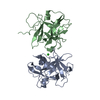


 PDBj
PDBj Assembly
 Mass: 18.015 Da / Num. of mol.: 197 / Source method: isolated from a natural source / Formula: H2O
 Sample preparation
 / Beamline: BL12-2 / Wavelength: 0.9184,0.9796,0.9795
 Processing