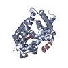
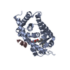
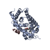
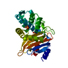
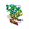
Entry Database : PDB / ID : 4ynkTitle Crystal structure of vitamin D receptor ligand binding domain complexed with a 19-norvitamin D compound Coactivator peptide drip from cDNA FLJ50196, highly similar to Peroxisome proliferator-activated receptor-binding protein Vitamin D3 receptor,Vitamin D3 receptor Keywords / Function / homology Function Domain/homology Component
/ / / / / / / / / / / / / / / / / / / / / / / / / / / / / / / / / / / / / / / / / / / / / / / / / / / / / / / / / / / / / / / / / / / / / / / / / / / / / / / / / / / / / / / / / / / / / / / / / / / / / / / / / / / / / / / / / / / / / / / / / / / / / / / / / / / / / / / / / / Biological species Rattus norvegicus (Norway rat)Homo sapiens (human)Method / / / Resolution : 2.3 Å Authors Watarai, Y. / Ikura, T. / Ito, N. Journal : J.Med.Chem. / Year : 2015Title : Synthesis, Biological Activities, and X-ray Crystal Structural Analysis of 25-Hydroxy-25(or 26)-adamantyl-17-[20(22),23-diynyl]-21-norvitamin D CompoundsAuthors : Watarai, Y. / Ishizawa, M. / Ikura, T. / Zacconi, F.C. / Uno, S. / Ito, N. / Mourino, A. / Tokiwa, H. / Makishima, M. / Yamada, S. History Deposition Mar 10, 2015 Deposition site / Processing site Revision 1.0 Jan 20, 2016 Provider / Type Revision 1.1 Feb 5, 2020 Group Data collection / Database references ... Data collection / Database references / Derived calculations / Refinement description Category citation / diffrn_source ... citation / diffrn_source / pdbx_struct_oper_list / refine Item _citation.journal_id_CSD / _diffrn_source.pdbx_synchrotron_site ... _citation.journal_id_CSD / _diffrn_source.pdbx_synchrotron_site / _pdbx_struct_oper_list.symmetry_operation / _refine.pdbx_method_to_determine_struct Revision 1.2 Nov 8, 2023 Group / Database references / Refinement descriptionCategory chem_comp_atom / chem_comp_bond ... chem_comp_atom / chem_comp_bond / database_2 / pdbx_initial_refinement_model Item / _database_2.pdbx_database_accession
Show all Show less
 Movie
Movie Controller

 Yorodumi
 Open data
 Basic information
 Components
 Keywords
 Function and homology information
 Homo sapiens (human)
 X-RAY DIFFRACTION /
  Authors
 Citation
 Structure visualization
 Downloads & links
 Other downloads


 PDBj
PDBj


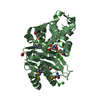
 Assembly

 Mass: 500.711 Da / Num. of mol.: 1 / Source method: obtained synthetically / Formula: C34H44O3
 Mass: 18.015 Da / Num. of mol.: 47 / Source method: isolated from a natural source / Formula: H2O
 Sample preparation
 / Beamline: BL38B1 / Wavelength: 1 Å
 Processing