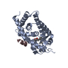
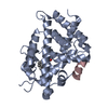
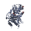
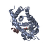
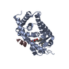
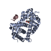
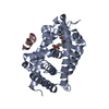
Entry Database : PDB / ID : 3vjsTitle Vitamin D receptor complex with a carborane compound Vitamin D3 receptor peptide from Mediator of RNA polymerase II transcription subunit 1 Keywords / / / Function / homology Function Domain/homology Component
/ / / / / / / / / / / / / / / / / / / / / / / / / / / / / / / / / / / / / / / / / / / / / / / / / / / / / / / / / / / / / / / / / / / / / / / / / / / / / / / / / / / / / / / / / / / / / / / / / / / / / / / / / / / / / / / / / / / / / / / / / / / / / / / / / / / / / / / / / / Biological species Rattus norvegicus (Norway rat)Homo sapiens (human)Method / / / Resolution : 1.93 Å Authors Fujii, S. / Masuno, M. / Kagechika, H. / Nakabayashi, M. / Ito, N. Journal : J.Am.Chem.Soc. / Year : 2011Title : Boron Cluster-based Development of Potent Nonsecosteroidal Vitamin D Receptor Ligands: Direct Observation of Hydrophobic Interaction between Protein Surface and CarboraneAuthors : Fujii, S. / Masuno, H. / Taoda, Y. / Kano, A. / Wongmayura, A. / Nakabayashi, M. / Ito, N. / Shimizu, M. / Kawachi, E. / Hirano, T. / Endo, Y. / Tanatani, A. / Kagechika, H. History Deposition Oct 31, 2011 Deposition site / Processing site Revision 1.0 Feb 8, 2012 Provider / Type Revision 1.1 Nov 8, 2023 Group Data collection / Database references ... Data collection / Database references / Derived calculations / Refinement description Category chem_comp_atom / chem_comp_bond ... chem_comp_atom / chem_comp_bond / database_2 / pdbx_initial_refinement_model / struct_ref_seq_dif / struct_site Item _database_2.pdbx_DOI / _database_2.pdbx_database_accession ... _database_2.pdbx_DOI / _database_2.pdbx_database_accession / _struct_ref_seq_dif.details / _struct_site.pdbx_auth_asym_id / _struct_site.pdbx_auth_comp_id / _struct_site.pdbx_auth_seq_id
Show all Show less
 Movie
Movie Controller


 Open data
 Basic information
 Components
 Keywords
 Function and homology information
 Homo sapiens (human)
 X-RAY DIFFRACTION /
  Authors
 Citation
 Structure visualization
 Downloads & links
 Other downloads
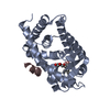


 PDBj
PDBj


 Assembly
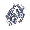

 Mass: 418.623 Da / Num. of mol.: 1 / Source method: obtained synthetically / Formula: C17H42B10O4
 Mass: 18.015 Da / Num. of mol.: 66 / Source method: isolated from a natural source / Formula: H2O
 Sample preparation
 / Beamline: BL-6A / Wavelength: 0.978 Å
 Processing