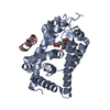
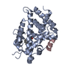
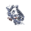
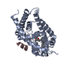
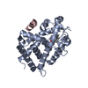
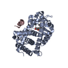
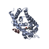
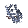
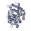
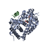
[English] 日本語
Yorodumi
- PDB-5zwe: Covalent bond formation between histidine of Vitamin D receptor (... -

+
Open data


ID or keywords:

Loading...

-
Basic information

Entry
Database: PDB / ID: 5zwe
TitleCovalent bond formation between histidine of Vitamin D receptor (VDR) and a full agonist having a vinyl ketone group via conjugate addition reaction
Components
  • 13-meric peptide from DRIP205 NR2 BOX peptide
  • Vitamin D3 receptor
KeywordsTRANSCRIPTION / HORMONE covalent modifier transcription factor VitaminD3 enone Michael addition electrophile
Function / homology
Function and homology information


enucleate erythrocyte development / positive regulation of type II interferon-mediated signaling pathway / mammary gland branching involved in thelarche / negative regulation of bone trabecula formation / Vitamin D (calciferol) metabolism / regulation of RNA biosynthetic process / androgen biosynthetic process / positive regulation of G0 to G1 transition / retinal pigment epithelium development / G0 to G1 transition ...enucleate erythrocyte development / positive regulation of type II interferon-mediated signaling pathway / mammary gland branching involved in thelarche / negative regulation of bone trabecula formation / Vitamin D (calciferol) metabolism / regulation of RNA biosynthetic process / androgen biosynthetic process / positive regulation of G0 to G1 transition / retinal pigment epithelium development / G0 to G1 transition / dense fibrillar component / SUMOylation of intracellular receptors / thyroid hormone mediated signaling pathway / Nuclear Receptor transcription pathway / bile acid nuclear receptor activity / response to bile acid / core mediator complex / regulation of vitamin D receptor signaling pathway / positive regulation of parathyroid hormone secretion / phosphate ion transmembrane transport / apoptotic process involved in mammary gland involution / vitamin D binding / cellular response to vitamin D / positive regulation of apoptotic process involved in mammary gland involution / calcitriol binding / lithocholic acid binding / mediator complex / nuclear retinoic acid receptor binding / ventricular trabecula myocardium morphogenesis / thyroid hormone generation / positive regulation of keratinocyte differentiation / Generic Transcription Pathway / embryonic heart tube development / negative regulation of ossification / positive regulation of hepatocyte proliferation / cellular response to thyroid hormone stimulus / positive regulation of vitamin D receptor signaling pathway / embryonic hindlimb morphogenesis / peroxisome proliferator activated receptor binding / nuclear vitamin D receptor binding / vitamin D receptor signaling pathway / bile acid signaling pathway / intestinal absorption / lens development in camera-type eye / nuclear thyroid hormone receptor binding / embryonic hemopoiesis / positive regulation of intracellular estrogen receptor signaling pathway / histone acetyltransferase binding / megakaryocyte development / erythrocyte development / response to aldosterone / cellular response to hepatocyte growth factor stimulus / cellular response to steroid hormone stimulus / epithelial cell proliferation involved in mammary gland duct elongation / LBD domain binding / fat cell differentiation / mammary gland branching involved in pregnancy / monocyte differentiation / regulation of calcium ion transport / decidualization / general transcription initiation factor binding / negative regulation of neuron differentiation / positive regulation of transcription initiation by RNA polymerase II / hematopoietic stem cell differentiation / embryonic placenta development / negative regulation of keratinocyte proliferation / animal organ regeneration / heterochromatin / ubiquitin ligase complex / nuclear retinoid X receptor binding / intracellular steroid hormone receptor signaling pathway / RNA polymerase II preinitiation complex assembly / keratinocyte differentiation / cellular response to epidermal growth factor stimulus / T-tubule / peroxisome proliferator activated receptor signaling pathway / RORA activates gene expression / lactation / Regulation of lipid metabolism by PPARalpha / BMAL1:CLOCK,NPAS2 activates circadian gene expression / nuclear receptor coactivator activity / Activation of gene expression by SREBF (SREBP) / positive regulation of erythrocyte differentiation / liver development / skeletal system development / nuclear estrogen receptor binding / promoter-specific chromatin binding / apoptotic signaling pathway / nuclear receptor binding / positive regulation of transcription elongation by RNA polymerase II / transcription coregulator activity / animal organ morphogenesis / brain development / euchromatin / Heme signaling / mRNA transcription by RNA polymerase II / cell morphogenesis / Transcriptional activation of mitochondrial biogenesis / transcription coactivator binding / intracellular calcium ion homeostasis
Similarity search - Function
Mediator complex, subunit Med1 / Mediator of RNA polymerase II transcription subunit 1 / Vitamin D receptor / VDR, DNA-binding domain / Retinoid X Receptor / Retinoid X Receptor / Nuclear hormone receptor / Nuclear hormones receptors DNA-binding region signature. / Zinc finger, nuclear hormone receptor-type / Zinc finger, C4 type (two domains) ...Mediator complex, subunit Med1 / Mediator of RNA polymerase II transcription subunit 1 / Vitamin D receptor / VDR, DNA-binding domain / Retinoid X Receptor / Retinoid X Receptor / Nuclear hormone receptor / Nuclear hormones receptors DNA-binding region signature. / Zinc finger, nuclear hormone receptor-type / Zinc finger, C4 type (two domains) / Nuclear hormone receptors DNA-binding domain profile. / c4 zinc finger in nuclear hormone receptors / Nuclear hormone receptor, ligand-binding domain / Nuclear hormone receptor-like domain superfamily / Ligand-binding domain of nuclear hormone receptor / Nuclear receptor (NR) ligand-binding (LBD) domain profile. / Ligand binding domain of hormone receptors / Zinc finger, NHR/GATA-type / Orthogonal Bundle / Mainly Alpha
Similarity search - Domain/homology
Chem-9K0 / Vitamin D3 receptor / Mediator of RNA polymerase II transcription subunit 1
Similarity search - Component
Biological speciesRattus norvegicus (Norway rat)
Homo sapiens (human)
MethodX-RAY DIFFRACTION / SYNCHROTRON / Resolution: 2.72 Å
AuthorsYoshizawa, M. / Itoh, T. / Anami, Y. / Kato, A. / Yoshimoto, N. / Yamamoto, K.
Funding support Japan, 1items
OrganizationGrant numberCountry
Japan
CitationJournal: J. Med. Chem. / Year: 2018
Title: Identification of the Histidine Residue in Vitamin D Receptor That Covalently Binds to Electrophilic Ligands
Authors: Yoshizawa, M. / Itoh, T. / Hori, T. / Kato, A. / Anami, Y. / Yoshimoto, N. / Yamamoto, K.
History
DepositionMay 15, 2018Deposition site: PDBJ / Processing site: PDBJ
Revision 1.0Jul 18, 2018Provider: repository / Type: Initial release
Revision 1.1Aug 8, 2018Group: Data collection / Database references / Category: citation / citation_author
Item: _citation.journal_volume / _citation.page_first ..._citation.journal_volume / _citation.page_first / _citation.page_last / _citation.title / _citation_author.identifier_ORCID

-
Structure visualization

Structure viewerMolecule:
MolmilJmol/JSmol

Downloads & links

-
Assembly

Deposited unit
A: Vitamin D3 receptor
C: 13-meric peptide from DRIP205 NR2 BOX peptide
hetero molecules


Theoretical massNumber of molelcules
Total (without water)32,5653
Polymers32,1662
Non-polymers3991
Water36020
1


  • Idetical with deposited unit
  • defined by author&software
  • Evidence: mass spectrometry
TypeNameSymmetry operationNumber
identity operation1_555x,y,z1
Buried area1160 Å2
ΔGint-11 kcal/mol
Surface area11580 Å2
MethodPISA
Unit cell
Length a, b, c (Å)153.530, 42.440, 41.870
Angle α, β, γ (deg.)90.00, 95.98, 90.00
Int Tables number5
Space group name H-MC121

-
Components

#1: Protein Vitamin D3 receptor / VDR / 1 / 25-dihydroxyvitamin D3 receptor / Nuclear receptor subfamily 1 group I member 1


Mass: 30595.037 Da / Num. of mol.: 1 / Mutation: 165-211 deletion
Source method: isolated from a genetically manipulated source
Source: (gene. exp.) Rattus norvegicus (Norway rat) / Gene: Vdr, Nr1i1 / Production host: Escherichia coli (E. coli) / References: UniProt: P13053
#2: Protein/peptide 13-meric peptide from DRIP205 NR2 BOX peptide


Mass: 1570.898 Da / Num. of mol.: 1
Source method: isolated from a genetically manipulated source
Source: (gene. exp.) Homo sapiens (human) / Production host: synthetic construct (others) / References: UniProt: Q15648*PLUS
#3: Chemical ChemComp-9K0 / (6R)-6-[(1R,3aS,4E,7aR)-7a-methyl-4-[2-[(3R,5R)-4-methylidene-3,5-bis(oxidanyl)cyclohexylidene]ethylidene]-2,3,3a,5,6,7-hexahydro-1H-inden-1-yl]hept-1-en-3-one


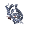
Mass: 398.578 Da / Num. of mol.: 1 / Source method: obtained synthetically / Formula: C26H38O3
#4: Water ChemComp-HOH / water / Water


Mass: 18.015 Da / Num. of mol.: 20 / Source method: isolated from a natural source / Formula: H2O

-
Experimental details

-
Experiment

ExperimentMethod: X-RAY DIFFRACTION / Number of used crystals: 1

-
Sample preparation

CrystalDensity Matthews: 2.11 Å3/Da / Density % sol: 41.67 %
Crystal growTemperature: 293 K / Method: vapor diffusion, hanging drop / Details: MOPS-Na, Na-Formate, PEG 4000, Ethyleneglycol

-
Data collection

DiffractionMean temperature: 100 K
Diffraction sourceSource: SYNCHROTRON / Site: Photon Factory / Beamline: BL-5A / Wavelength: 1 Å
DetectorType: ADSC QUANTUM 315r / Detector: CCD / Date: Nov 15, 2015
RadiationProtocol: SINGLE WAVELENGTH / Monochromatic (M) / Laue (L): M / Scattering type: x-ray
Radiation wavelengthWavelength: 1 Å / Relative weight: 1
ReflectionResolution: 2.72→41.64 Å / Num. obs: 7161 / % possible obs: 94.7 % / Redundancy: 3.6 % / Net I/σ(I): 88.8
Reflection shellResolution: 2.72→2.86 Å

-
Processing

Software
NameVersionClassification
REFMAC5.8.0103refinement
MOSFLMdata reduction
SCALAdata scaling
REFMACphasing
RefinementResolution: 2.72→41.64 Å / Cor.coef. Fo:Fc: 0.919 / Cor.coef. Fo:Fc free: 0.9 / Cross valid method: THROUGHOUT / ESU R Free: 0.405 / Details: HYDROGENS HAVE BEEN ADDED IN THE RIDING POSITIONS
RfactorNum. reflection% reflectionSelection details
Rfree0.2519 693 9.7 %RANDOM
Rwork0.22585 ---
obs0.22839 6463 96.94 %-
Solvent computationIon probe radii: 0.8 Å / Shrinkage radii: 0.8 Å / VDW probe radii: 1.2 Å
Displacement parametersBiso mean: 40.228 Å2
Baniso -1Baniso -2Baniso -3
1--3.9 Å2-0 Å2-1.43 Å2
2--5.63 Å2-0 Å2
3----1.41 Å2
Refinement stepCycle: 1 / Resolution: 2.72→41.64 Å
ProteinNucleic acidLigandSolventTotal
Num. atoms1981 0 29 20 2030
Refine LS restraints
Refine-IDTypeDev idealDev ideal targetNumber
X-RAY DIFFRACTIONr_bond_refined_d0.0270.0192085
X-RAY DIFFRACTIONr_bond_other_d00.022031
X-RAY DIFFRACTIONr_angle_refined_deg1.2211.9952823
X-RAY DIFFRACTIONr_angle_other_deg3.97534695
X-RAY DIFFRACTIONr_dihedral_angle_1_deg4.0895253
X-RAY DIFFRACTIONr_dihedral_angle_2_deg34.34124.59887
X-RAY DIFFRACTIONr_dihedral_angle_3_deg17.87415375
X-RAY DIFFRACTIONr_dihedral_angle_4_deg20.876159
X-RAY DIFFRACTIONr_chiral_restr0.0710.2324
X-RAY DIFFRACTIONr_gen_planes_refined0.0110.0212281
X-RAY DIFFRACTIONr_gen_planes_other0.030.02444
X-RAY DIFFRACTIONr_nbd_refined
X-RAY DIFFRACTIONr_nbd_other
X-RAY DIFFRACTIONr_nbtor_refined
X-RAY DIFFRACTIONr_nbtor_other
X-RAY DIFFRACTIONr_xyhbond_nbd_refined
X-RAY DIFFRACTIONr_xyhbond_nbd_other
X-RAY DIFFRACTIONr_metal_ion_refined
X-RAY DIFFRACTIONr_metal_ion_other
X-RAY DIFFRACTIONr_symmetry_vdw_refined
X-RAY DIFFRACTIONr_symmetry_vdw_other
X-RAY DIFFRACTIONr_symmetry_hbond_refined
X-RAY DIFFRACTIONr_symmetry_hbond_other
X-RAY DIFFRACTIONr_symmetry_metal_ion_refined
X-RAY DIFFRACTIONr_symmetry_metal_ion_other
X-RAY DIFFRACTIONr_mcbond_it1.5554.0761015
X-RAY DIFFRACTIONr_mcbond_other1.5464.0751013
X-RAY DIFFRACTIONr_mcangle_it2.5076.1021267
X-RAY DIFFRACTIONr_mcangle_other2.5076.1021268
X-RAY DIFFRACTIONr_scbond_it1.5584.141070
X-RAY DIFFRACTIONr_scbond_other1.5584.141070
X-RAY DIFFRACTIONr_scangle_it
X-RAY DIFFRACTIONr_scangle_other2.4416.171557
X-RAY DIFFRACTIONr_long_range_B_refined4.43338.0318919
X-RAY DIFFRACTIONr_long_range_B_other4.42938.0328914
X-RAY DIFFRACTIONr_rigid_bond_restr
X-RAY DIFFRACTIONr_sphericity_free
X-RAY DIFFRACTIONr_sphericity_bonded
LS refinement shellResolution: 2.724→2.795 Å / Total num. of bins used: 20
RfactorNum. reflection% reflection
Rfree0.336 40 -
Rwork0.285 410 -
obs--82.42 %

+
About Yorodumi

-
News

-
Feb 9, 2022. New format data for meta-information of EMDB entries

New format data for meta-information of EMDB entries

  • Version 3 of the EMDB header file is now the official format.
  • The previous official version 1.9 will be removed from the archive.

Related info.:EMDB header

External links:wwPDB to switch to version 3 of the EMDB data model

-
Aug 12, 2020. Covid-19 info

Covid-19 info

URL: https://pdbj.org/emnavi/covid19.php

New page: Covid-19 featured information page in EM Navigator.

Related info.:Covid-19 info / Mar 5, 2020. Novel coronavirus structure data

+
Mar 5, 2020. Novel coronavirus structure data

Novel coronavirus structure data

Related info.:Yorodumi Speices / Aug 12, 2020. Covid-19 info

External links:COVID-19 featured content - PDBj / Molecule of the Month (242):Coronavirus Proteases

+
Jan 31, 2019. EMDB accession codes are about to change! (news from PDBe EMDB page)

EMDB accession codes are about to change! (news from PDBe EMDB page)

  • The allocation of 4 digits for EMDB accession codes will soon come to an end. Whilst these codes will remain in use, new EMDB accession codes will include an additional digit and will expand incrementally as the available range of codes is exhausted. The current 4-digit format prefixed with “EMD-” (i.e. EMD-XXXX) will advance to a 5-digit format (i.e. EMD-XXXXX), and so on. It is currently estimated that the 4-digit codes will be depleted around Spring 2019, at which point the 5-digit format will come into force.
  • The EM Navigator/Yorodumi systems omit the EMD- prefix.

Related info.:Q: What is EMD? / ID/Accession-code notation in Yorodumi/EM Navigator

External links:EMDB Accession Codes are Changing Soon! / Contact to PDBj

+
Jul 12, 2017. Major update of PDB

Major update of PDB

  • wwPDB released updated PDB data conforming to the new PDBx/mmCIF dictionary.
  • This is a major update changing the version number from 4 to 5, and with Remediation, in which all the entries are updated.
  • In this update, many items about electron microscopy experimental information are reorganized (e.g. em_software).
  • Now, EM Navigator and Yorodumi are based on the updated data.

External links:wwPDB Remediation / Enriched Model Files Conforming to OneDep Data Standards Now Available in the PDB FTP Archive

-
Yorodumi

Thousand views of thousand structures

  • Yorodumi is a browser for structure data from EMDB, PDB, SASBDB, etc.
  • This page is also the successor to EM Navigator detail page, and also detail information page/front-end page for Omokage search.
  • The word "yorodu" (or yorozu) is an old Japanese word meaning "ten thousand". "mi" (miru) is to see.

Related info.:EMDB / PDB / SASBDB / Comparison of 3 databanks / Yorodumi Search / Aug 31, 2016. New EM Navigator & Yorodumi / Yorodumi Papers / Jmol/JSmol / Function and homology information / Changes in new EM Navigator and Yorodumi

Read more