 Movie
Movie Controller

[English] 日本語
 Yorodumi
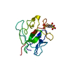
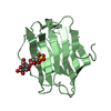
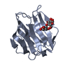
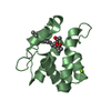
- PDB-4ym0: Crystal structure of the human galectin-4 C-terminal carbohydrate... -
+ Open data

- Basic information
| Entry | Database: PDB / ID: 4ym0 | |||||||||
|---|---|---|---|---|---|---|---|---|---|---|
| Title | Crystal structure of the human galectin-4 C-terminal carbohydrate recognition domain in complex with lacto-N-tetraose (LNT) | |||||||||
 Components Components | Galectin-4 | |||||||||
 Keywords Keywords | SUGAR BINDING PROTEIN / galectin / lectin / carbohydrate binding protein / lacto-N-tetraose / carbohydrate recognition / beta sandwich | |||||||||
| Function / homology |  Function and homology information Function and homology informationantibacterial peptide biosynthetic process / galactoside binding / carbohydrate binding / extracellular matrix / cell adhesion / : / plasma membrane / cytosol Similarity search - Function | |||||||||
| Biological species |  Homo sapiens (human) Homo sapiens (human) | |||||||||
| Method |  X-RAY DIFFRACTION / SYNCHROTRON / MOLECULAR REPLACEMENT / Resolution: 2.3 Å X-RAY DIFFRACTION / SYNCHROTRON / MOLECULAR REPLACEMENT / Resolution: 2.3 Å | |||||||||
 Authors Authors | Bum-Erdene, K. / Blanchard, H. | |||||||||
| Funding support |  Australia, 1items Australia, 1items
| |||||||||
 Citation Citation | Journal: Febs J. / Year: 2015 Title: Structural characterization of human galectin-4 C-terminal domain: elucidating the molecular basis for recognition of glycosphingolipids, sulfated saccharides and blood group antigens. Authors: Bum-Erdene, K. / Leffler, H. / Nilsson, U.J. / Blanchard, H. | |||||||||
| History |
|
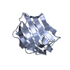
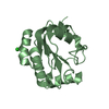
- Structure visualization
| Structure viewer | Molecule: MolmilJmol/JSmol |
|---|
- Downloads & links
-Download
| PDBx/mmCIF format | 4ym0.cif.gz | 126.5 KB | Display | PDBx/mmCIF format |
|---|---|---|---|---|
| PDB format | pdb4ym0.ent.gz | 97.9 KB | Display | PDB format |
| PDBx/mmJSON format | 4ym0.json.gz | Tree view | PDBx/mmJSON format | |
| Others |  Other downloads Other downloads |
-Validation report
| Arichive directory | https://data.pdbj.org/pub/pdb/validation_reports/ym/4ym0ftp://data.pdbj.org/pub/pdb/validation_reports/ym/4ym0 | HTTPS FTP |
|---|
-Related structure data


-Links
 PDBj
PDBj


- Assembly
| Deposited unit | 
| ||||||||
|---|---|---|---|---|---|---|---|---|---|
| 1 | 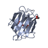
| ||||||||
| 2 | 
| ||||||||
| 3 | 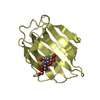
| ||||||||
| 4 | 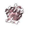
| ||||||||
| Unit cell |
|
-Components
-Protein , 1 types, 4 molecules ABDC
| #1: Protein | Mass: 16962.291 Da / Num. of mol.: 4 / Fragment: unp residues 171-323 Source method: isolated from a genetically manipulated source Source: (gene. exp.) Homo sapiens (human) / Gene: LGALS4 / Production host:  |
|---|
-Sugars , 3 types, 4 molecules
| #2: Polysaccharide | beta-D-galactopyranose-(1-3)-2-acetamido-2-deoxy-beta-D-glucopyranose Source method: isolated from a genetically manipulated source | ||
|---|---|---|---|
| #3: Polysaccharide | Source method: isolated from a genetically manipulated source #4: Polysaccharide | beta-D-galactopyranose-(1-4)-beta-D-glucopyranose / beta-lactose |   Source method: isolated from a genetically manipulated source Details: oligosaccharide / References: beta-lactose |
-Non-polymers , 2 types, 129 molecules 

| #5: Chemical | ChemComp-GOL /  Mass: 92.094 Da / Num. of mol.: 1 / Source method: obtained synthetically / Formula: C3H8O3 Mass: 92.094 Da / Num. of mol.: 1 / Source method: obtained synthetically / Formula: C3H8O3 |
|---|---|
| #6: Water | ChemComp-HOH / Mass: 18.015 Da / Num. of mol.: 128 / Source method: isolated from a natural source / Formula: H2O |
-Experimental details
-Experiment
| Experiment | Method: X-RAY DIFFRACTION / Number of used crystals: 1 |
|---|
- Sample preparation
| Crystal | Density Matthews: 1.96 Å3/Da / Density % sol: 37.18 % |
|---|---|
| Crystal grow | Temperature: 293.15 K / Method: vapor diffusion, hanging drop / pH: 7 Details: 1.2 M ammonium sulfate, 0.1 M HEPES pH 7.0, 4 % v/v PEG 400 PH range: 7 |
-Data collection
| Diffraction | Mean temperature: 100 K | |||||||||||||||||||||||||||
|---|---|---|---|---|---|---|---|---|---|---|---|---|---|---|---|---|---|---|---|---|---|---|---|---|---|---|---|---|
| Diffraction source | Source: SYNCHROTRON / Site: Australian Synchrotron / Beamline: MX1 / Wavelength: 0.9537 Å | |||||||||||||||||||||||||||
| Detector | Type: ADSC QUANTUM 210r / Detector: CCD / Date: Oct 15, 2013 | |||||||||||||||||||||||||||
| Radiation | Protocol: SINGLE WAVELENGTH / Monochromatic (M) / Laue (L): M / Scattering type: x-ray | |||||||||||||||||||||||||||
| Radiation wavelength | Wavelength: 0.9537 Å / Relative weight: 1 | |||||||||||||||||||||||||||
| Reflection twin |
| |||||||||||||||||||||||||||
| Reflection | Resolution: 2.3→44.99 Å / Num. obs: 22849 / % possible obs: 98.6 % / Redundancy: 4.1 % / CC1/2: 0.999 / Rmerge(I) obs: 0.028 / Rpim(I) all: 0.016 / Net I/σ(I): 35.2 / Num. measured all: 93135 | |||||||||||||||||||||||||||
| Reflection shell | Diffraction-ID: 1 / Rejects: _
|
- Processing
| Software |
| |||||||||||||||||||||||||||||||||||||||||||||||||||||||||||||||||||||||||||
|---|---|---|---|---|---|---|---|---|---|---|---|---|---|---|---|---|---|---|---|---|---|---|---|---|---|---|---|---|---|---|---|---|---|---|---|---|---|---|---|---|---|---|---|---|---|---|---|---|---|---|---|---|---|---|---|---|---|---|---|---|---|---|---|---|---|---|---|---|---|---|---|---|---|---|---|---|
| Refinement | Method to determine structure: MOLECULAR REPLACEMENT / Resolution: 2.3→42.54 Å / Cor.coef. Fo:Fc: 0.946 / Cor.coef. Fo:Fc free: 0.938 / WRfactor Rfree: 0.197 / WRfactor Rwork: 0.1641 / FOM work R set: 0.8138 / SU B: 7.17 / SU ML: 0.178 / SU R Cruickshank DPI: 0.0989 / SU Rfree: 0.0475 / Cross valid method: THROUGHOUT / σ(F): 0 / ESU R: 0.099 / ESU R Free: 0.048 / Stereochemistry target values: MAXIMUM LIKELIHOOD Details: HYDROGENS HAVE BEEN ADDED IN THE RIDING POSITIONS U VALUES : REFINED INDIVIDUALLY
| |||||||||||||||||||||||||||||||||||||||||||||||||||||||||||||||||||||||||||
| Solvent computation | Ion probe radii: 0.8 Å / Shrinkage radii: 0.8 Å / VDW probe radii: 1.2 Å / Solvent model: MASK | |||||||||||||||||||||||||||||||||||||||||||||||||||||||||||||||||||||||||||
| Displacement parameters | Biso max: 68.15 Å2 / Biso mean: 28.378 Å2 / Biso min: 9.64 Å2
| |||||||||||||||||||||||||||||||||||||||||||||||||||||||||||||||||||||||||||
| Refinement step | Cycle: final / Resolution: 2.3→42.54 Å
| |||||||||||||||||||||||||||||||||||||||||||||||||||||||||||||||||||||||||||
| Refine LS restraints |
| |||||||||||||||||||||||||||||||||||||||||||||||||||||||||||||||||||||||||||
| LS refinement shell | Resolution: 2.297→2.356 Å / Total num. of bins used: 20
|
