| Entry | Database: PDB / ID: 4yia
|
|---|
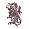
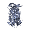
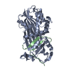
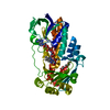
| Title | Structural mechanism of hormone release in thyroxine binding globulin |
|---|
 Components Components | (Thyroxine-binding ...) x 2 |
|---|
 Keywords Keywords | SIGNALING PROTEIN / Thyroxine Binding globulin / Thyroxine / Serpin / hormone release / cation Pi interaction |
|---|
| Function / homology |  Function and homology information Function and homology information
thyroid hormone transport / serine-type endopeptidase inhibitor activity / : / extracellular exosome / extracellular regionSimilarity search - Function Antithrombin; Chain I, domain 2 / Antithrombin, subunit I, domain 2 / Alpha-1-antitrypsin; domain 1 / Alpha-1-antitrypsin, domain 1 / Serpin, conserved site / Serpins signature. / Serpin superfamily, domain 2 / Serpin family / Serpin domain / Serpin superfamily ...Antithrombin; Chain I, domain 2 / Antithrombin, subunit I, domain 2 / Alpha-1-antitrypsin; domain 1 / Alpha-1-antitrypsin, domain 1 / Serpin, conserved site / Serpins signature. / Serpin superfamily, domain 2 / Serpin family / Serpin domain / Serpin superfamily / Serpin superfamily, domain 1 / Serpin (serine protease inhibitor) / SERine Proteinase INhibitors / Roll / 2-Layer Sandwich / Mainly Beta / Alpha BetaSimilarity search - Domain/homology |
|---|
| Biological species |  Homo sapiens (human) Homo sapiens (human) |
|---|
| Method |  X-RAY DIFFRACTION / SYNCHROTRON / MOLECULAR REPLACEMENT / Resolution: 1.58 Å X-RAY DIFFRACTION / SYNCHROTRON / MOLECULAR REPLACEMENT / Resolution: 1.58 Å |
|---|
 Authors Authors | Zheng, Y. |
|---|
| Funding support |  China, 1items China, 1items | Organization | Grant number | Country |
|---|
| | China |
|
|---|
 Citation Citation | Journal: To Be Published
Title: Structural mechanism of hormone release in thyroxine Binding globulin
Authors: Zheng, Y. |
|---|
| History | | Deposition | Mar 1, 2015 | Deposition site: RCSB / Processing site: PDBJ |
|---|
| Revision 1.0 | Mar 2, 2016 | Provider: repository / Type: Initial release |
|---|
| Revision 1.1 | Mar 20, 2024 | Group: Data collection / Database references / Derived calculations
Category: chem_comp_atom / chem_comp_bond ...chem_comp_atom / chem_comp_bond / database_2 / pdbx_struct_conn_angle / pdbx_struct_oper_list / pdbx_struct_special_symmetry / struct_conn
Item: _database_2.pdbx_DOI / _database_2.pdbx_database_accession ..._database_2.pdbx_DOI / _database_2.pdbx_database_accession / _pdbx_struct_conn_angle.ptnr1_auth_comp_id / _pdbx_struct_conn_angle.ptnr1_auth_seq_id / _pdbx_struct_conn_angle.ptnr1_label_asym_id / _pdbx_struct_conn_angle.ptnr1_label_atom_id / _pdbx_struct_conn_angle.ptnr1_label_comp_id / _pdbx_struct_conn_angle.ptnr1_label_seq_id / _pdbx_struct_conn_angle.ptnr1_symmetry / _pdbx_struct_conn_angle.ptnr3_auth_comp_id / _pdbx_struct_conn_angle.ptnr3_auth_seq_id / _pdbx_struct_conn_angle.ptnr3_label_asym_id / _pdbx_struct_conn_angle.ptnr3_label_atom_id / _pdbx_struct_conn_angle.ptnr3_label_comp_id / _pdbx_struct_conn_angle.ptnr3_label_seq_id / _pdbx_struct_conn_angle.ptnr3_symmetry / _pdbx_struct_conn_angle.value / _pdbx_struct_oper_list.symmetry_operation / _struct_conn.pdbx_dist_value / _struct_conn.ptnr1_auth_asym_id / _struct_conn.ptnr1_auth_comp_id / _struct_conn.ptnr1_auth_seq_id / _struct_conn.ptnr1_label_asym_id / _struct_conn.ptnr1_label_atom_id / _struct_conn.ptnr1_label_comp_id / _struct_conn.ptnr1_label_seq_id / _struct_conn.ptnr2_auth_asym_id / _struct_conn.ptnr2_auth_comp_id / _struct_conn.ptnr2_auth_seq_id / _struct_conn.ptnr2_label_asym_id / _struct_conn.ptnr2_label_atom_id / _struct_conn.ptnr2_label_comp_id / _struct_conn.ptnr2_symmetry |
|---|
|
|---|
 Movie
Movie Controller

 Yorodumi
 Open data
 Basic information
 Structure visualization
 Downloads & links
 Other downloads
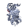


 PDBj
PDBj

 Assembly


 Mass: 40.078 Da / Num. of mol.: 2 / Source method: obtained synthetically / Formula: Ca
 Mass: 22.990 Da / Num. of mol.: 2 / Source method: obtained synthetically / Formula: Na
 Mass: 35.453 Da / Num. of mol.: 2 / Source method: obtained synthetically / Formula: Cl
 Mass: 357.788 Da / Num. of mol.: 1 / Source method: obtained synthetically / Formula: C19H16ClNO4 / Comment: medication, antiinflammatory*YM
 Sample preparation
 Processing