 Movie
Movie Controller


+ Open data

- Basic information
| Entry | Database: PDB / ID: 4yg7 | ||||||
|---|---|---|---|---|---|---|---|
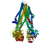
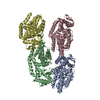
| Title | Structure of FL autorepression promoter complex | ||||||
 Components Components |
| ||||||
 Keywords Keywords | TRANSCRIPTION / TRANSFERASE/DNA / persistence / multidrug tolerance / autorepression / promoter / TRANSFERASE-DNA complex | ||||||
| Function / homology |  Function and homology information Function and homology informationdormancy process / toxin-antitoxin complex / single-species biofilm formation / regulation of growth / DNA-binding transcription repressor activity / core promoter sequence-specific DNA binding / protein-DNA complex / sequence-specific DNA binding / non-specific serine/threonine protein kinase / transcription cis-regulatory region binding ...dormancy process / toxin-antitoxin complex / single-species biofilm formation / regulation of growth / DNA-binding transcription repressor activity / core promoter sequence-specific DNA binding / protein-DNA complex / sequence-specific DNA binding / non-specific serine/threonine protein kinase / transcription cis-regulatory region binding / response to antibiotic / protein serine kinase activity / negative regulation of DNA-templated transcription / protein serine/threonine kinase activity / DNA-templated transcription / regulation of DNA-templated transcription / magnesium ion binding / protein homodimerization activity / ATP binding / cytosol Similarity search - Function | ||||||
| Biological species |  | ||||||
| Method |  X-RAY DIFFRACTION / SYNCHROTRON / MOLECULAR REPLACEMENT / Resolution: 3.77 Å X-RAY DIFFRACTION / SYNCHROTRON / MOLECULAR REPLACEMENT / Resolution: 3.77 Å | ||||||
 Authors Authors | Schumacher, M.A. | ||||||
 Citation Citation | Journal: Nature / Year: 2015 Title: HipBA-promoter structures reveal the basis of heritable multidrug tolerance. Authors: Schumacher, M.A. / Balani, P. / Min, J. / Chinnam, N.B. / Hansen, S. / Vulic, M. / Lewis, K. / Brennan, R.G. | ||||||
| History |
|
- Structure visualization
| Structure viewer | Molecule: MolmilJmol/JSmol |
|---|
- Downloads & links
-Download
| PDBx/mmCIF format | 4yg7.cif.gz | 282.6 KB | Display | PDBx/mmCIF format |
|---|---|---|---|---|
| PDB format | pdb4yg7.ent.gz | 223.1 KB | Display | PDB format |
| PDBx/mmJSON format | 4yg7.json.gz | Tree view | PDBx/mmJSON format | |
| Others |  Other downloads Other downloads |
-Validation report
| Summary document | 4yg7_validation.pdf.gz | 498.3 KB | Display | wwPDB validaton report |
|---|---|---|---|---|
| Full document | 4yg7_full_validation.pdf.gz | 642.8 KB | Display | |
| Data in XML | 4yg7_validation.xml.gz | 62 KB | Display | |
| Data in CIF | 4yg7_validation.cif.gz | 84.2 KB | Display | |
| Arichive directory | https://data.pdbj.org/pub/pdb/validation_reports/yg/4yg7ftp://data.pdbj.org/pub/pdb/validation_reports/yg/4yg7 | HTTPS FTP |
-Related structure data
| Related structure data |  4yg1C 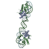 4yg4C 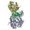 5k98C  3dnvS C: citing same article ( S: Starting model for refinement |
|---|---|
| Similar structure data |


-Links
 PDBj
PDBj


- Assembly
| Deposited unit | 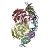
| ||||||||
|---|---|---|---|---|---|---|---|---|---|
| 1 |
| ||||||||
| Unit cell |
|
-Components
| #1: Protein | Mass: 8060.239 Da / Num. of mol.: 4 / Fragment: UNP residues 4-74 Source method: isolated from a genetically manipulated source Source: (gene. exp.) Strain: K12 / Gene: hipB, b1508, JW1501 / Production host: #2: Protein | Mass: 48946.203 Da / Num. of mol.: 2 Source method: isolated from a genetically manipulated source Source: (gene. exp.) Strain: K12 / Gene: hipA, b1507, JW1500 / Production host: References: UniProt: P23874, non-specific serine/threonine protein kinase #3: DNA chain | | Mass: 15398.923 Da / Num. of mol.: 1 / Source method: obtained synthetically / Source: (synth.) #4: DNA chain | | Mass: 15414.922 Da / Num. of mol.: 1 / Source method: obtained synthetically / Source: (synth.) |
|---|
-Experimental details
-Experiment
| Experiment | Method: X-RAY DIFFRACTION / Number of used crystals: 1 |
|---|
- Sample preparation
| Crystal | Density Matthews: 5.29 Å3/Da / Density % sol: 76.75 % |
|---|---|
| Crystal grow | Temperature: 298 K / Method: vapor diffusion, hanging drop / Details: sodium formate / PH range: 5-7 |
-Data collection
| Diffraction | Mean temperature: 100 K |
|---|---|
| Diffraction source | Source: SYNCHROTRON / Site: ALS  / Beamline: 8.3.1 / Wavelength: 1 Å / Beamline: 8.3.1 / Wavelength: 1 Å |
| Detector | Type: ADSC QUANTUM 315r / Detector: CCD / Date: Sep 23, 2013 |
| Radiation | Monochromator: double crystal Si(111) / Protocol: SINGLE WAVELENGTH / Monochromatic (M) / Laue (L): M / Scattering type: x-ray |
| Radiation wavelength | Wavelength: 1 Å / Relative weight: 1 |
| Reflection | Resolution: 3.77→161.4 Å / Num. obs: 34997 / % possible obs: 63.6 % / Redundancy: 4 % / Net I/σ(I): 4 |
| Reflection shell | Mean I/σ(I) obs: 1.4 / Rsym value: 0.762 |
- Processing
| Software |
| ||||||||||||||||||||||||||||
|---|---|---|---|---|---|---|---|---|---|---|---|---|---|---|---|---|---|---|---|---|---|---|---|---|---|---|---|---|---|
| Refinement | Method to determine structure: MOLECULAR REPLACEMENT Starting model: PDB entry 3DNV Resolution: 3.77→161.4 Å / Cross valid method: THROUGHOUT / σ(F): 0 Details: AUTHOR STATES ONLY MINIMAL REFINEMENT WAS PERFORMED.
| ||||||||||||||||||||||||||||
| Solvent computation | Bsol: 72.4475 Å2 | ||||||||||||||||||||||||||||
| Displacement parameters | Biso max: 267.11 Å2 / Biso mean: 180.4669 Å2 / Biso min: 1 Å2
| ||||||||||||||||||||||||||||
| Refinement step | Cycle: final / Resolution: 3.77→161.4 Å
| ||||||||||||||||||||||||||||
| Refine LS restraints |
| ||||||||||||||||||||||||||||
| LS refinement shell | Highest resolution: 3.77 Å / Rfactor Rfree: 0.46 | ||||||||||||||||||||||||||||
| Xplor file |
|
