 Movie
Movie Controller

[English] 日本語
 Yorodumi
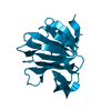
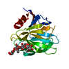
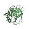
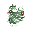
- PDB-4yg3: Structural basis of glycan recognition in neonate-specific rotaviruses -
+ Open data

- Basic information
| Entry | Database: PDB / ID: 4yg3 | ||||||
|---|---|---|---|---|---|---|---|
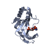
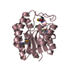
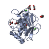
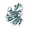
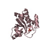
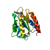
| Title | Structural basis of glycan recognition in neonate-specific rotaviruses | ||||||
 Components Components | Outer capsid protein VP4 | ||||||
 Keywords Keywords | VIRAL PROTEIN / rotavirus / structural biology / glycan | ||||||
| Function / homology |  Function and homology information Function and homology informationhost cell rough endoplasmic reticulum / permeabilization of host organelle membrane involved in viral entry into host cell / host cytoskeleton / viral outer capsid / host cell endoplasmic reticulum-Golgi intermediate compartment / virion attachment to host cell / host cell plasma membrane Similarity search - Function | ||||||
| Biological species |  Rotavirus A Rotavirus A | ||||||
| Method |  X-RAY DIFFRACTION / SYNCHROTRON / MOLECULAR REPLACEMENT / Resolution: 2.285 Å X-RAY DIFFRACTION / SYNCHROTRON / MOLECULAR REPLACEMENT / Resolution: 2.285 Å | ||||||
 Authors Authors | Hu, L. / Prasad, B.V.V. | ||||||
| Funding support |  United States, 1items United States, 1items
| ||||||
 Citation Citation | Journal: Nat Commun / Year: 2015 Title: Structural basis of glycan specificity in neonate-specific bovine-human reassortant rotavirus. Authors: Hu, L. / Ramani, S. / Czako, R. / Sankaran, B. / Yu, Y. / Smith, D.F. / Cummings, R.D. / Estes, M.K. / Venkataram Prasad, B.V. | ||||||
| History |
|
- Structure visualization
| Structure viewer | Molecule: MolmilJmol/JSmol |
|---|
- Downloads & links
-Download
| PDBx/mmCIF format | 4yg3.cif.gz | 48.8 KB | Display | PDBx/mmCIF format |
|---|---|---|---|---|
| PDB format | pdb4yg3.ent.gz | 32.4 KB | Display | PDB format |
| PDBx/mmJSON format | 4yg3.json.gz | Tree view | PDBx/mmJSON format | |
| Others |  Other downloads Other downloads |
-Validation report
| Arichive directory | https://data.pdbj.org/pub/pdb/validation_reports/yg/4yg3ftp://data.pdbj.org/pub/pdb/validation_reports/yg/4yg3 | HTTPS FTP |
|---|
-Related structure data
| Related structure data |  4yfwSC  4yfzC 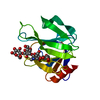 4yg0C 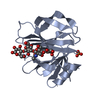 4yg6C  4yez S: Starting model for refinement C: citing same article ( |
|---|---|
| Similar structure data |

-Links
 PDBj
PDBj- Assembly
| Deposited unit | 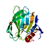
| ||||||||
|---|---|---|---|---|---|---|---|---|---|
| 1 |
| ||||||||
| Unit cell |
|
-Components
| #1: Protein | Mass: 18063.570 Da / Num. of mol.: 1 / Fragment: UNP residues 65-225 Source method: isolated from a genetically manipulated source Source: (gene. exp.) Rotavirus A / Production host:  | ||
|---|---|---|---|
| #2: Chemical |   Mass: 96.063 Da / Num. of mol.: 3 / Source method: obtained synthetically / Formula: SO4 Mass: 96.063 Da / Num. of mol.: 3 / Source method: obtained synthetically / Formula: SO4#3: Water | ChemComp-HOH / |  Mass: 18.015 Da / Num. of mol.: 81 / Source method: isolated from a natural source / Formula: H2O Mass: 18.015 Da / Num. of mol.: 81 / Source method: isolated from a natural source / Formula: H2O |
-Experimental details
-Experiment
| Experiment | Method: X-RAY DIFFRACTION / Number of used crystals: 1 |
|---|
- Sample preparation
| Crystal | Density Matthews: 1.72 Å3/Da / Density % sol: 28.55 % |
|---|---|
| Crystal grow | Temperature: 293 K / Method: vapor diffusion, hanging drop / pH: 8 / Details: 0.1 M Tris, 1.6 M Ammonium sulfate |
-Data collection
| Diffraction | Mean temperature: 100 K |
|---|---|
| Diffraction source | Source: SYNCHROTRON / Site: ALS / Beamline: 5.0.1 / Wavelength: 0.977408 Å |
| Detector | Type: ADSC QUANTUM 315r / Detector: CCD / Date: Jan 24, 2014 |
| Radiation | Monochromator: Asymmetric curved crystal / Protocol: SINGLE WAVELENGTH / Monochromatic (M) / Laue (L): M / Scattering type: x-ray |
| Radiation wavelength | Wavelength: 0.977408 Å / Relative weight: 1 |
| Reflection | Resolution: 2.28→30 Å / Num. obs: 5508 / % possible obs: 99.2 % / Redundancy: 3.7 % / Rmerge(I) obs: 0.112 / Net I/σ(I): 10.8 |
| Reflection shell | Resolution: 2.28→2.32 Å / Redundancy: 3.6 % / Rmerge(I) obs: 0.223 / Mean I/σ(I) obs: 5.8 / % possible all: 99.6 |
- Processing
| Software |
| ||||||||||||||||||||||||
|---|---|---|---|---|---|---|---|---|---|---|---|---|---|---|---|---|---|---|---|---|---|---|---|---|---|
| Refinement | Method to determine structure: MOLECULAR REPLACEMENT Starting model: 4YFW Resolution: 2.285→29.643 Å / SU ML: 0.24 / Cross valid method: FREE R-VALUE / σ(F): 1.37 / Phase error: 24.62 / Stereochemistry target values: ML
| ||||||||||||||||||||||||
| Solvent computation | Shrinkage radii: 0.9 Å / VDW probe radii: 1.11 Å / Solvent model: FLAT BULK SOLVENT MODEL | ||||||||||||||||||||||||
| Refinement step | Cycle: LAST / Resolution: 2.285→29.643 Å
| ||||||||||||||||||||||||
| Refine LS restraints |
| ||||||||||||||||||||||||
| LS refinement shell |
|
