- PDB-4yeh: Crystal structure of Mg2+ ion containing hemopexin fold from Kabu... -
+
Open data
ID or keywords:
Loading...
-
Basic information
Entry
Database: PDB / ID: 4yeh
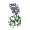
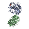
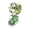
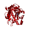
Title
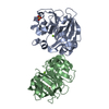
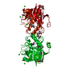
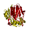
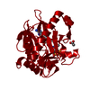
Crystal structure of Mg2+ ion containing hemopexin fold from Kabuli chana (chickpea white) at 2.45A resolution reveals a structural basis of metal ion transport
Resolution: 2.45→69.7 Å / Cor.coef. Fo:Fc: 0.935 / Cor.coef. Fo:Fc free: 0.892 / SU B: 7.194 / SU ML: 0.167 / Cross valid method: THROUGHOUT / ESU R: 0.119 / ESU R Free: 0.049 / Stereochemistry target values: MAXIMUM LIKELIHOOD / Details: HYDROGENS HAVE BEEN USED IF PRESENT IN THE INPUT
Rfactor
Num. reflection
% reflection
Selection details
Rfree
0.19388
890
5 %
RANDOM
Rwork
0.15066
-
-
-
obs
0.15284
17005
96.96 %
-
Solvent computation
Ion probe radii: 0.8 Å / Shrinkage radii: 0.8 Å / VDW probe radii: 1.2 Å / Solvent model: MASK
 Movie
Movie Controller

 Yorodumi
 Open data
 Basic information
 Components
 Keywords
 Function and homology information
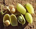 Cicer arietinum (chickpea)
 X-RAY DIFFRACTION /
  Authors
 Citation
 Structure visualization
 Downloads & links
 Other downloads


 PDBj
PDBj
 Assembly
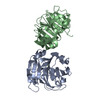

 Mass: 24.305 Da / Num. of mol.: 2 / Source method: obtained synthetically / Formula: Mg
 Mass: 18.015 Da / Num. of mol.: 222 / Source method: isolated from a natural source / Formula: H2O
 Sample preparation
 / Beamline: BM14 / Wavelength: 0.97 Å
 Processing