Resolution: 2.2→19.94 Å / Cor.coef. Fo:Fc: 0.955 / Cor.coef. Fo:Fc free: 0.925 / SU B: 11.09 / SU ML: 0.129 / Cross valid method: THROUGHOUT / ESU R: 0.283 / ESU R Free: 0.207 / Stereochemistry target values: MAXIMUM LIKELIHOOD / Details: HYDROGENS HAVE BEEN USED IF PRESENT IN THE INPUT
Rfactor
Num. reflection
% reflection
Selection details
Rfree
0.2258
1214
5.1 %
RANDOM
Rwork
0.17909
-
-
-
obs
0.18155
22431
99.34 %
-
all
-
23739
-
-
Solvent computation
Ion probe radii: 0.8 Å / Shrinkage radii: 0.8 Å / VDW probe radii: 1.2 Å / Solvent model: MASK
Displacement parameters
Biso mean: 26.404 Å2
Baniso -1
Baniso -2
Baniso -3
1-
-0.05 Å2
-0 Å2
0 Å2
2-
-
0.04 Å2
-0 Å2
3-
-
-
0.01 Å2
Refine analyze
Luzzati coordinate error obs: 0.2161 Å / Luzzati d res low obs: 19.93 Å
Refinement step
Cycle: LAST / Resolution: 2.2→19.94 Å
Protein
Nucleic acid
Ligand
Solvent
Total
Num. atoms
3590
0
19
121
3730
Refine LS restraints
Refine-ID
Type
Dev ideal
Dev ideal target
Number
X-RAY DIFFRACTION
r_bond_refined_d
0.009
0.02
3694
X-RAY DIFFRACTION
r_angle_refined_deg
1.227
1.954
4986
X-RAY DIFFRACTION
r_dihedral_angle_1_deg
6.234
5
446
X-RAY DIFFRACTION
r_dihedral_angle_2_deg
27.469
23.444
180
X-RAY DIFFRACTION
r_dihedral_angle_3_deg
16.393
15
602
X-RAY DIFFRACTION
r_dihedral_angle_4_deg
12.863
15
20
X-RAY DIFFRACTION
r_chiral_restr
0.091
0.2
512
X-RAY DIFFRACTION
r_gen_planes_refined
0.005
0.021
2866
LS refinement shell
Resolution: 2.201→2.258 Å / Total num. of bins used: 20
Rfactor
Num. reflection
% reflection
Rfree
0.232
70
-
Rwork
0.191
1385
-
obs
-
1512
93.39 %
Refinement TLS params.
Method: refined / Refine-ID: X-RAY DIFFRACTION
ID
L11 (°2)
L12 (°2)
L13 (°2)
L22 (°2)
L23 (°2)
L33 (°2)
S11 (Å °)
S12 (Å °)
S13 (Å °)
S21 (Å °)
S22 (Å °)
S23 (Å °)
S31 (Å °)
S32 (Å °)
S33 (Å °)
T11 (Å2)
T12 (Å2)
T13 (Å2)
T22 (Å2)
T23 (Å2)
T33 (Å2)
Origin x (Å)
Origin y (Å)
Origin z (Å)
1
0.6925
0.0195
-0.1086
0.2397
0.1591
0.2977
0.0201
0.0228
-0.0157
0.014
-0.0079
0.0057
0.0083
0.0217
-0.0122
0.0435
0.0065
0.0036
0.0075
-0.0033
0.0278
48.2662
18.1569
9.3085
2
0.9008
0.1127
-0.3799
0.1574
-0.2965
0.7353
0.036
-0.0218
-0.0189
-0.0048
-0.0119
-0.0073
0.0024
-0.0347
-0.0241
0.034
0.0004
0.0097
0.0254
0.0176
0.0153
17.3224
18.1152
34.0761
Refinement TLS group
ID
Refine-ID
Refine TLS-ID
Auth asym-ID
Auth seq-ID
1
X-RAY DIFFRACTION
1
A
4 - 301
2
X-RAY DIFFRACTION
2
B
4 - 285
+
About Yorodumi
-
News
-
Feb 9, 2022. New format data for meta-information of EMDB entries
New format data for meta-information of EMDB entries
Version 3 of the EMDB header file is now the official format.
The previous official version 1.9 will be removed from the archive.
In the structure databanks used in Yorodumi, some data are registered as the other names, "COVID-19 virus" and "2019-nCoV". Here are the details of the virus and the list of structure data.
Jan 31, 2019. EMDB accession codes are about to change! (news from PDBe EMDB page)
EMDB accession codes are about to change! (news from PDBe EMDB page)
The allocation of 4 digits for EMDB accession codes will soon come to an end. Whilst these codes will remain in use, new EMDB accession codes will include an additional digit and will expand incrementally as the available range of codes is exhausted. The current 4-digit format prefixed with “EMD-” (i.e. EMD-XXXX) will advance to a 5-digit format (i.e. EMD-XXXXX), and so on. It is currently estimated that the 4-digit codes will be depleted around Spring 2019, at which point the 5-digit format will come into force.
The EM Navigator/Yorodumi systems omit the EMD- prefix.
Related info.:Q: What is EMD? / ID/Accession-code notation in Yorodumi/EM Navigator
Yorodumi is a browser for structure data from EMDB, PDB, SASBDB, etc.
This page is also the successor to EM Navigator detail page, and also detail information page/front-end page for Omokage search.
The word "yorodu" (or yorozu) is an old Japanese word meaning "ten thousand". "mi" (miru) is to see.
Related info.:EMDB / PDB / SASBDB / Comparison of 3 databanks / Yorodumi Search / Aug 31, 2016. New EM Navigator & Yorodumi / Yorodumi Papers / Jmol/JSmol / Function and homology information / Changes in new EM Navigator and Yorodumi
 Movie
Movie Controller

 Yorodumi
 Open data
 Basic information
 Components
 Keywords
 Function and homology information
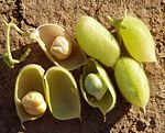 Cicer arietinum (chickpea)
 X-RAY DIFFRACTION /
  Authors
 Citation
 Structure visualization
 Downloads & links
 Other downloads


 PDBj
PDBj
 Assembly


 Mass: 126.904 Da / Num. of mol.: 2 / Source method: obtained synthetically / Formula: I
 Mass: 40.078 Da / Num. of mol.: 2 / Source method: obtained synthetically / Formula: Ca
 Mass: 35.453 Da / Num. of mol.: 3 / Source method: obtained synthetically / Formula: Cl
 Mass: 22.990 Da / Num. of mol.: 2 / Source method: obtained synthetically / Formula: Na
 Mass: 96.063 Da / Num. of mol.: 2 / Source method: obtained synthetically / Formula: SO4
 Sample preparation
 Processing