Source: ROTATING ANODE / Type: BRUKER AXS MICROSTAR / Wavelength: 1.5418 Å
Detector
Type: MAR scanner 345 mm plate / Detector: IMAGE PLATE / Date: Sep 26, 2011 / Details: Mirrors
Radiation
Monochromator: OSMIC MIRROR / Protocol: SINGLE WAVELENGTH / Monochromatic (M) / Laue (L): M / Scattering type: x-ray
Radiation wavelength
Wavelength: 1.5418 Å / Relative weight: 1
Reflection
Redundancy: 6.1 % / Av σ(I) over netI: 7 / Number: 95010 / Rsym value: 0.095 / D res high: 1.881 Å / D res low: 76.89 Å / Num. obs: 15680 / % possible obs: 96.4
Diffraction reflection shell
Highest resolution (Å)
Lowest resolution (Å)
% possible obs (%)
ID
Rmerge(I) obs
Rsym value
Redundancy
2.09
2.2
81.6
1
0.497
0.497
5.6
2.2
2.33
98
1
0.364
0.364
6
2.33
2.49
98.2
1
0.284
0.284
6.1
2.49
2.69
98.5
1
0.211
0.211
6.2
2.69
2.95
99.2
1
0.133
0.133
6.2
2.95
3.3
99.3
1
0.096
0.096
6.2
3.3
3.81
99.6
1
0.064
0.064
6.2
3.81
4.66
99.7
1
0.053
0.053
6.1
4.66
6.59
99.8
1
0.05
0.05
6
6.59
54.84
99.7
1
0.032
0.032
5.5
Reflection
Resolution: 2.09→54.84 Å / Num. obs: 15680 / % possible obs: 96.4 % / Redundancy: 6.1 % / Biso Wilson estimate: 27.2 Å2 / Rmerge(I) obs: 0.095 / Net I/σ(I): 12.4
Reflection shell
Resolution: 2.09→2.2 Å / Redundancy: 5.6 % / Rmerge(I) obs: 0.497 / Mean I/σ(I) obs: 3.4 / Num. unique all: 10732 / % possible all: 81.6
Resolution: 2.1→54.84 Å / Cor.coef. Fo:Fc: 0.942 / Cor.coef. Fo:Fc free: 0.922 / WRfactor Rfree: 0.2276 / WRfactor Rwork: 0.1855 / FOM work R set: 0.8401 / SU B: 5.05 / SU ML: 0.136 / SU R Cruickshank DPI: 0.2313 / SU Rfree: 0.1915 / Cross valid method: THROUGHOUT / ESU R: 0.231 / ESU R Free: 0.192 / Stereochemistry target values: MAXIMUM LIKELIHOOD Details: HYDROGENS HAVE BEEN USED IF PRESENT IN THE INPUT U VALUES: REFINED INDIVIDUALLY
Rfactor
Num. reflection
% reflection
Selection details
Rfree
0.24394
828
5.3 %
RANDOM
Rwork
0.20113
-
-
-
obs
0.2034
14846
98.03 %
-
Solvent computation
Ion probe radii: 0.8 Å / Shrinkage radii: 0.8 Å / VDW probe radii: 1.2 Å / Solvent model: MASK
 Movie
Movie Controller


 Open data
 Basic information
 Components
 Keywords
 Function and homology information
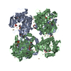
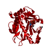
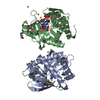
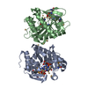
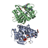
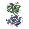
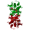
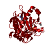
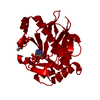
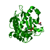
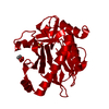
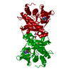
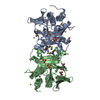
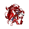
 Pyrococcus horikoshii (archaea)
 X-RAY DIFFRACTION /
  Authors
 Citation
 Structure visualization
 Downloads & links
 Other downloads


 PDBj
PDBj Assembly
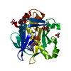


 Mass: 427.201 Da / Num. of mol.: 1 / Source method: obtained synthetically / Formula: C10H15N5O10P2 / Comment: ADP, energy-carrying molecule*YM
 Mass: 94.971 Da / Num. of mol.: 2 / Source method: obtained synthetically / Formula: PO4
 Mass: 59.044 Da / Num. of mol.: 1 / Source method: obtained synthetically / Formula: C2H3O2
 Mass: 24.305 Da / Num. of mol.: 1 / Source method: obtained synthetically / Formula: Mg
 Sample preparation
 Processing