[English] 日本語
Yorodumi
- PDB-4ydn: High resolution crystal structure of human transthyretin bound to... -

+
Open data


ID or keywords:

Loading...

-
Basic information

Entry
Database: PDB / ID: 4ydn
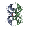
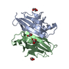
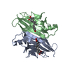
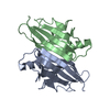
Title
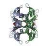
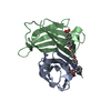
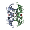
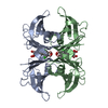
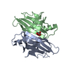
High resolution crystal structure of human transthyretin bound to ligand and conjugates of 4-(5-(3,5-dichloro-4-hydroxyphenyl)-1,3,4-oxadiazol-2-yl)phenyl fluorosulfate
ComponentsTransthyretin
KeywordsTRANSPORT PROTEIN / Hormone transport protein / Thyroxine / retinol
Function / homology
Function and homology information


Retinoid cycle disease events / The canonical retinoid cycle in rods (twilight vision) / thyroid hormone binding / purine nucleobase metabolic process / Non-integrin membrane-ECM interactions / Retinoid metabolism and transport / hormone activity / azurophil granule lumen / Amyloid fiber formation / Neutrophil degranulation ...Retinoid cycle disease events / The canonical retinoid cycle in rods (twilight vision) / thyroid hormone binding / purine nucleobase metabolic process / Non-integrin membrane-ECM interactions / Retinoid metabolism and transport / hormone activity / azurophil granule lumen / Amyloid fiber formation / Neutrophil degranulation / extracellular space / extracellular exosome / extracellular region / identical protein binding
Similarity search - Function
Transthyretin/hydroxyisourate hydrolase domain / Transthyretin, conserved site / Transthyretin signature 2. / Transthyretin, thyroxine binding site / Transthyretin signature 1. / Transthyretin / Transthyretin/hydroxyisourate hydrolase / Transthyretin/hydroxyisourate hydrolase domain / Transthyretin/hydroxyisourate hydrolase domain superfamily / HIUase/Transthyretin family ...Transthyretin/hydroxyisourate hydrolase domain / Transthyretin, conserved site / Transthyretin signature 2. / Transthyretin, thyroxine binding site / Transthyretin signature 1. / Transthyretin / Transthyretin/hydroxyisourate hydrolase / Transthyretin/hydroxyisourate hydrolase domain / Transthyretin/hydroxyisourate hydrolase domain superfamily / HIUase/Transthyretin family / Immunoglobulin-like / Sandwich / Mainly Beta
Similarity search - Domain/homology
Chem-2WN / Chem-4B8 / Transthyretin
Similarity search - Component
Biological speciesHomo sapiens (human)
MethodX-RAY DIFFRACTION / SYNCHROTRON / MOLECULAR REPLACEMENT / Resolution: 1.35 Å
AuthorsConnelly, S. / Bradbury, N.C.
CitationJournal: J.Am.Chem.Soc. / Year: 2015
Title: A Fluorogenic Aryl Fluorosulfate for Intraorganellar Transthyretin Imaging in Living Cells and in Caenorhabditis elegans.
Authors: Baranczak, A. / Liu, Y. / Connelly, S. / Du, W.G. / Greiner, E.R. / Genereux, J.C. / Wiseman, R.L. / Eisele, Y.S. / Bradbury, N.C. / Dong, J. / Noodleman, L. / Sharpless, K.B. / Wilson, I.A. ...Authors: Baranczak, A. / Liu, Y. / Connelly, S. / Du, W.G. / Greiner, E.R. / Genereux, J.C. / Wiseman, R.L. / Eisele, Y.S. / Bradbury, N.C. / Dong, J. / Noodleman, L. / Sharpless, K.B. / Wilson, I.A. / Encalada, S.E. / Kelly, J.W.
History
DepositionFeb 22, 2015Deposition site: RCSB / Processing site: RCSB
Revision 1.0Jul 15, 2015Provider: repository / Type: Initial release
Revision 1.1Nov 1, 2017Group: Advisory / Author supporting evidence ...Advisory / Author supporting evidence / Derived calculations / Refinement description / Source and taxonomy
Category: entity_src_gen / pdbx_struct_assembly_auth_evidence ...entity_src_gen / pdbx_struct_assembly_auth_evidence / pdbx_struct_oper_list / pdbx_validate_close_contact / software
Item: _entity_src_gen.pdbx_alt_source_flag / _pdbx_struct_oper_list.symmetry_operation / _software.classification
Revision 1.2Sep 27, 2023Group: Data collection / Database references ...Data collection / Database references / Derived calculations / Refinement description
Category: chem_comp_atom / chem_comp_bond ...chem_comp_atom / chem_comp_bond / database_2 / pdbx_initial_refinement_model / pdbx_struct_special_symmetry
Item: _database_2.pdbx_DOI / _database_2.pdbx_database_accession

-
Structure visualization

Structure viewerMolecule:
MolmilJmol/JSmol

Downloads & links

-
Assembly

Deposited unit
A: Transthyretin
B: Transthyretin
hetero molecules


Theoretical massNumber of molelcules
Total (without water)28,8465
Polymers27,7132
Non-polymers1,1343
Water2,810156
1
A: Transthyretin
B: Transthyretin
hetero molecules

A: Transthyretin
B: Transthyretin
hetero molecules


Theoretical massNumber of molelcules
Total (without water)57,69310
Polymers55,4264
Non-polymers2,2676
Water724
TypeNameSymmetry operationNumber
identity operation1_555x,y,z1
crystal symmetry operation2_555-x,-y,z1
Buried area6190 Å2
ΔGint16.5 kcal/mol
Surface area19490 Å2
MethodPISA
Unit cell
Length a, b, c (Å)43.093, 85.522, 64.650
Angle α, β, γ (deg.)90.000, 90.000, 90.000
Int Tables number18
Space group name H-MP21212
Components on special symmetry positions
IDModelComponents
11A-201-

2WN

21A-201-

2WN

31A-201-

2WN

41A-201-

2WN

51A-202-

4B8

61A-202-

4B8

71A-202-

4B8

81B-201-

2WN

91B-201-

2WN

101B-201-

2WN

111B-201-

2WN

121B-201-

2WN

DetailsTetramer confirmed by by size exclusion chromatography

-
Components

#1: Protein Transthyretin / ATTR / Prealbumin / TBPA


Mass: 13856.417 Da / Num. of mol.: 2 / Fragment: UNP residues 21-147
Source method: isolated from a genetically manipulated source
Source: (gene. exp.) Homo sapiens (human) / Gene: TTR, PALB / Plasmid: pmmHA / Production host: Escherichia coli (E. coli) / References: UniProt: P02766
#2: Chemical ChemComp-2WN / 4-[5-(3,5-dichloro-4-hydroxyphenyl)-1,3,4-oxadiazol-2-yl]phenyl sulfurofluoridate


Mass: 405.185 Da / Num. of mol.: 2 / Source method: obtained synthetically / Formula: C14H7Cl2FN2O5S
#3: Chemical ChemComp-4B8 / 2,6-dichloro-4-[5-(4-hydroxyphenyl)-1,3,4-oxadiazol-2-yl]phenol


Mass: 323.131 Da / Num. of mol.: 1 / Source method: obtained synthetically / Formula: C14H8Cl2N2O3
#4: Water ChemComp-HOH / water


Mass: 18.015 Da / Num. of mol.: 156 / Source method: isolated from a natural source / Formula: H2O

-
Experimental details

-
Experiment

ExperimentMethod: X-RAY DIFFRACTION / Number of used crystals: 1

-
Sample preparation

CrystalDensity Matthews: 2.33 Å3/Da / Density % sol: 42.77 %
Crystal growTemperature: 298 K / Method: vapor diffusion, sitting drop / pH: 5.5
Details: The wt-TTR was concentrated to 4 mg/ml in 10 mM NaPi, 100 mM KCl, at pH 7.6 and co-crystallized at room temperature with inhibitors using the vapor-diffusion sitting drop method, crystals ...Details: The wt-TTR was concentrated to 4 mg/ml in 10 mM NaPi, 100 mM KCl, at pH 7.6 and co-crystallized at room temperature with inhibitors using the vapor-diffusion sitting drop method, crystals were grown from 1.395 M sodium citrate, 3.5% v/v glycerol at ph 5.5. The crystals were frozen using a cryo-protectant solution of 1.395 m sodium citrate, ph 5.5, containing 10% v/v glycerol, vapor diffusion, sitting drop, temperature 298.0

-
Data collection

DiffractionMean temperature: 100 K
Diffraction sourceSource: SYNCHROTRON / Site: SSRL / Beamline: BL11-1 / Wavelength: 0.97945 Å
DetectorType: PSI PILATUS 6M / Detector: PIXEL / Date: Nov 22, 2013
RadiationMonochromator: ASYMMETRIC CUT 4.965 DEGS SIDE SCATTERING BENT CUBE-ROOT I -BEAM SINGLE CRYSTAL
Protocol: SINGLE WAVELENGTH / Monochromatic (M) / Laue (L): M / Scattering type: x-ray
Radiation wavelengthWavelength: 0.97945 Å / Relative weight: 1
ReflectionResolution: 1.349→38.484 Å / Num. all: 53165 / Num. obs: 53165 / % possible obs: 99.5 % / Redundancy: 6.4 % / Biso Wilson estimate: 14.6 Å2 / Rpim(I) all: 0.012 / Rrim(I) all: 0.031 / Rsym value: 0.028 / Net I/av σ(I): 13.804 / Net I/σ(I): 28.4 / Num. measured all: 338134
Reflection shell

Diffraction-ID: 1 / Rejects: _

Resolution (Å)Redundancy (%)Rmerge(I) obsMean I/σ(I) obsNum. measured allNum. unique allRpim(I) allRsym valueNet I/σ(I) obs% possible all
1.35-1.426.20.4491.64678375140.1940.4494.898
1.42-1.516.30.2762.64588572590.1190.2767.399.8
1.51-1.616.60.1554.64527568750.0650.15511.999.9
1.61-1.746.20.0997.23981263980.0430.09916.999.8
1.74-1.916.70.05612.73946359050.0230.05626.9100
1.91-2.136.30.03618.93393053610.0160.03640.499.8
2.13-2.466.60.02923.43122847600.0120.02951.499.8
2.46-3.026.20.02426.42506740430.0110.02459.199.8
3.02-4.276.20.01932.51990431970.0080.01974.399.4
4.27-38.4845.80.01731.61078718530.0080.01773.799.5

-
Processing

Software
NameVersionClassification
SCALA3.3.21data scaling
REFMAC5.8.0049refinement
PDB_EXTRACT3.15data extraction
XDSdata scaling
XDSdata reduction
RefinementMethod to determine structure: MOLECULAR REPLACEMENT
Starting model: 2QGB
Resolution: 1.35→38.484 Å / Cor.coef. Fo:Fc: 0.973 / Cor.coef. Fo:Fc free: 0.959 / WRfactor Rfree: 0.1917 / WRfactor Rwork: 0.1581 / FOM work R set: 0.8886 / SU B: 1.625 / SU ML: 0.031 / SU R Cruickshank DPI: 0.0572 / SU Rfree: 0.0556 / Cross valid method: THROUGHOUT / σ(F): 0 / ESU R: 0.057 / ESU R Free: 0.056 / Stereochemistry target values: MAXIMUM LIKELIHOOD
Details: HYDROGENS HAVE BEEN ADDED IN THE RIDING POSITIONS U VALUES : REFINED INDIVIDUALLY
RfactorNum. reflection% reflectionSelection details
Rfree0.1951 2693 5 %RANDOM
Rwork0.1574 50399 --
obs0.1591 50399 99.4 %-
Solvent computationIon probe radii: 0.8 Å / Shrinkage radii: 0.8 Å / VDW probe radii: 1.2 Å / Solvent model: MASK
Displacement parametersBiso max: 91.06 Å2 / Biso mean: 21.614 Å2 / Biso min: 10.04 Å2
Baniso -1Baniso -2Baniso -3
1-0.13 Å20 Å20 Å2
2---0.32 Å20 Å2
3---0.19 Å2
Refinement stepCycle: final / Resolution: 1.35→38.484 Å
ProteinNucleic acidLigandSolventTotal
Num. atoms1789 0 96 156 2041
Biso mean--23.09 33.46 -
Num. residues----231
Refine LS restraints
Refine-IDTypeDev idealDev ideal targetNumber
X-RAY DIFFRACTIONr_bond_refined_d0.0150.022082
X-RAY DIFFRACTIONr_bond_other_d0.0020.021866
X-RAY DIFFRACTIONr_angle_refined_deg1.6942.0132877
X-RAY DIFFRACTIONr_angle_other_deg1.3613.0034335
X-RAY DIFFRACTIONr_dihedral_angle_1_deg5.9355268
X-RAY DIFFRACTIONr_dihedral_angle_2_deg30.79123.64785
X-RAY DIFFRACTIONr_dihedral_angle_3_deg13.76915311
X-RAY DIFFRACTIONr_dihedral_angle_4_deg15.4811511
X-RAY DIFFRACTIONr_chiral_restr0.0820.2311
X-RAY DIFFRACTIONr_gen_planes_refined0.0090.0212364
X-RAY DIFFRACTIONr_gen_planes_other0.0060.02471
X-RAY DIFFRACTIONr_mcbond_it2.0921.805974
X-RAY DIFFRACTIONr_mcbond_other2.0131.801973
X-RAY DIFFRACTIONr_mcangle_it2.4172.7081229
X-RAY DIFFRACTIONr_rigid_bond_restr3.01233948
X-RAY DIFFRACTIONr_sphericity_free20.602555
X-RAY DIFFRACTIONr_sphericity_bonded8.12853974
LS refinement shellResolution: 1.349→1.384 Å / Total num. of bins used: 20
RfactorNum. reflection% reflection
Rfree0.233 180 -
Rwork0.213 3531 -
all-3711 -
obs--95.99 %

+
About Yorodumi

-
News

-
Feb 9, 2022. New format data for meta-information of EMDB entries

New format data for meta-information of EMDB entries

  • Version 3 of the EMDB header file is now the official format.
  • The previous official version 1.9 will be removed from the archive.

Related info.:EMDB header

External links:wwPDB to switch to version 3 of the EMDB data model

-
Aug 12, 2020. Covid-19 info

Covid-19 info

URL: https://pdbjlc1.pdbj.org/emnavi/covid19.php

New page: Covid-19 featured information page in EM Navigator.

Related info.:Covid-19 info / Mar 5, 2020. Novel coronavirus structure data

+
Mar 5, 2020. Novel coronavirus structure data

Novel coronavirus structure data

Related info.:Yorodumi Speices / Aug 12, 2020. Covid-19 info

External links:COVID-19 featured content - PDBj / Molecule of the Month (242):Coronavirus Proteases

+
Jan 31, 2019. EMDB accession codes are about to change! (news from PDBe EMDB page)

EMDB accession codes are about to change! (news from PDBe EMDB page)

  • The allocation of 4 digits for EMDB accession codes will soon come to an end. Whilst these codes will remain in use, new EMDB accession codes will include an additional digit and will expand incrementally as the available range of codes is exhausted. The current 4-digit format prefixed with “EMD-” (i.e. EMD-XXXX) will advance to a 5-digit format (i.e. EMD-XXXXX), and so on. It is currently estimated that the 4-digit codes will be depleted around Spring 2019, at which point the 5-digit format will come into force.
  • The EM Navigator/Yorodumi systems omit the EMD- prefix.

Related info.:Q: What is EMD? / ID/Accession-code notation in Yorodumi/EM Navigator

External links:EMDB Accession Codes are Changing Soon! / Contact to PDBj

+
Jul 12, 2017. Major update of PDB

Major update of PDB

  • wwPDB released updated PDB data conforming to the new PDBx/mmCIF dictionary.
  • This is a major update changing the version number from 4 to 5, and with Remediation, in which all the entries are updated.
  • In this update, many items about electron microscopy experimental information are reorganized (e.g. em_software).
  • Now, EM Navigator and Yorodumi are based on the updated data.

External links:wwPDB Remediation / Enriched Model Files Conforming to OneDep Data Standards Now Available in the PDB FTP Archive

-
Yorodumi

Thousand views of thousand structures

  • Yorodumi is a browser for structure data from EMDB, PDB, SASBDB, etc.
  • This page is also the successor to EM Navigator detail page, and also detail information page/front-end page for Omokage search.
  • The word "yorodu" (or yorozu) is an old Japanese word meaning "ten thousand". "mi" (miru) is to see.

Related info.:EMDB / PDB / SASBDB / Comparison of 3 databanks / Yorodumi Search / Aug 31, 2016. New EM Navigator & Yorodumi / Yorodumi Papers / Jmol/JSmol / Function and homology information / Changes in new EM Navigator and Yorodumi

Read more