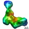
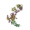
Entry Database : PDB / ID : 4ydhTitle The structure of human FMNL1 N-terminal domains bound to Cdc42 Cell division control protein 42 homolog Formin-like protein 1 Keywords / / / Function / homology Function Domain/homology Component
/ / / / / / / / / / / / / / / / / / / / / / / / / / / / / / / / / / / / / / / / / / / / / / / / / / / / / / / / / / / / / / / / / / / / / / / / / / / / / / / / / / / / / / / / / / / / / / / / / / / / / / / / / / / / / / / / / / / / / / / / / / / / / / / / / / / / / / / / / / / / / / / / / / / / Biological species Homo sapiens (human)Method / / / Resolution : 3.8 Å Authors Kuhn, S. / Anand, K. / Geyer, M. Journal : Nat Commun / Year : 2015Title : The structure of FMNL2-Cdc42 yields insights into the mechanism of lamellipodia and filopodia formation.Authors : Kuhn, S. / Erdmann, C. / Kage, F. / Block, J. / Schwenkmezger, L. / Steffen, A. / Rottner, K. / Geyer, M. History Deposition Feb 22, 2015 Deposition site / Processing site Revision 1.0 May 13, 2015 Provider / Type Revision 1.1 Feb 20, 2019 Group / Data collection / Derived calculationsCategory pdbx_data_processing_status / pdbx_validate_close_contact ... pdbx_data_processing_status / pdbx_validate_close_contact / struct_conn / struct_conn_type Revision 1.2 Jan 10, 2024 Group Data collection / Database references ... Data collection / Database references / Derived calculations / Refinement description Category chem_comp_atom / chem_comp_bond ... chem_comp_atom / chem_comp_bond / database_2 / pdbx_initial_refinement_model / struct_conn Item _database_2.pdbx_DOI / _database_2.pdbx_database_accession ... _database_2.pdbx_DOI / _database_2.pdbx_database_accession / _struct_conn.pdbx_dist_value / _struct_conn.ptnr1_auth_asym_id / _struct_conn.ptnr1_auth_comp_id / _struct_conn.ptnr1_auth_seq_id / _struct_conn.ptnr1_label_asym_id / _struct_conn.ptnr1_label_atom_id / _struct_conn.ptnr1_label_comp_id / _struct_conn.ptnr1_label_seq_id / _struct_conn.ptnr2_auth_asym_id / _struct_conn.ptnr2_label_asym_id
Show all Show less
 Movie
Movie Controller


 Open data
 Basic information
 Components
 Keywords
 Function and homology information
 Homo sapiens (human)
 X-RAY DIFFRACTION /
  Authors
 Citation
 Structure visualization
 Downloads & links
 Other downloads


 PDBj
PDBj


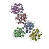
 Assembly
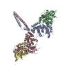


 Mass: 522.196 Da / Num. of mol.: 2 / Source method: obtained synthetically / Formula: C10H17N6O13P3
 Mass: 24.305 Da / Num. of mol.: 2 / Source method: obtained synthetically / Formula: Mg
 Sample preparation
 / Beamline: X06SA / Wavelength: 0.9786 Å
 Processing