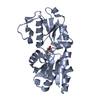
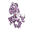
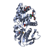
 Movie
Movie Controller


+ Open data

- Basic information
| Entry | Database: PDB / ID: 4yah | ||||||
|---|---|---|---|---|---|---|---|
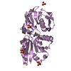
| Title | Crystal Structure of the Methionine Binding Protein, MetQ | ||||||
 Components Components | D-methionine-binding lipoprotein MetQ | ||||||
 Keywords Keywords | TRANSPORT PROTEIN / Solute binding protein / Methionine Transport | ||||||
| Function / homology |  Function and homology information Function and homology informationmethionine-importing ABC transporter complex / methionine import across plasma membrane / D-methionine transmembrane transport / methionine transport / ATP-binding cassette (ABC) transporter complex, substrate-binding subunit-containing / outer membrane-bounded periplasmic space / membrane / plasma membrane Similarity search - Function | ||||||
| Biological species |  | ||||||
| Method |  X-RAY DIFFRACTION / SYNCHROTRON / MOLECULAR REPLACEMENT / molecular replacement / Resolution: 1.6 Å X-RAY DIFFRACTION / SYNCHROTRON / MOLECULAR REPLACEMENT / molecular replacement / Resolution: 1.6 Å | ||||||
 Authors Authors | Lai, J.Y. / Kadaba, N.S. / Kaiser, J.T. / Rees, D.C. | ||||||
 Citation Citation | Journal: Biol.Chem. / Year: 2015 Title: The contribution of methionine to the stability of the Escherichia coli MetNIQ ABC transporter-substrate binding protein complex. Authors: Nguyen, P.T. / Li, Q.W. / Kadaba, N.S. / Lai, J.Y. / Yang, J.G. / Rees, D.C. | ||||||
| History |
|
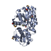
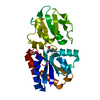
- Structure visualization
| Structure viewer | Molecule: MolmilJmol/JSmol |
|---|
- Downloads & links
-Download
| PDBx/mmCIF format | 4yah.cif.gz | 111.9 KB | Display | PDBx/mmCIF format |
|---|---|---|---|---|
| PDB format | pdb4yah.ent.gz | 84.9 KB | Display | PDB format |
| PDBx/mmJSON format | 4yah.json.gz | Tree view | PDBx/mmJSON format | |
| Others |  Other downloads Other downloads |
-Validation report
| Summary document | 4yah_validation.pdf.gz | 424.1 KB | Display | wwPDB validaton report |
|---|---|---|---|---|
| Full document | 4yah_full_validation.pdf.gz | 424.1 KB | Display | |
| Data in XML | 4yah_validation.xml.gz | 14.6 KB | Display | |
| Data in CIF | 4yah_validation.cif.gz | 22.5 KB | Display | |
| Arichive directory | https://data.pdbj.org/pub/pdb/validation_reports/ya/4yahftp://data.pdbj.org/pub/pdb/validation_reports/ya/4yah | HTTPS FTP |
-Related structure data
| Related structure data |  1p99S S: Starting model for refinement |
|---|---|
| Similar structure data |


-Links
 PDBj
PDBj


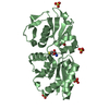
- Assembly
| Deposited unit | 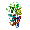
| ||||||||
|---|---|---|---|---|---|---|---|---|---|
| 1 |
| ||||||||
| Unit cell |
|
-Components
| #1: Protein | Mass: 30534.701 Da / Num. of mol.: 1 Source method: isolated from a genetically manipulated source Source: (gene. exp.) Strain: K12 / Gene: metQ, yaeC, b0197, JW0193 / Plasmid: pET21b+ / Production host: |
|---|---|
| #2: Chemical | ChemComp-MET /   Type: L-peptide linking / Mass: 149.211 Da / Num. of mol.: 1 / Source method: obtained synthetically / Formula: C5H11NO2S Type: L-peptide linking / Mass: 149.211 Da / Num. of mol.: 1 / Source method: obtained synthetically / Formula: C5H11NO2S |
| #3: Water | ChemComp-HOH /  Mass: 18.015 Da / Num. of mol.: 359 / Source method: isolated from a natural source / Formula: H2O Mass: 18.015 Da / Num. of mol.: 359 / Source method: isolated from a natural source / Formula: H2O |
-Experimental details
-Experiment
| Experiment | Method: X-RAY DIFFRACTION / Number of used crystals: 1 |
|---|
- Sample preparation
| Crystal | Density Matthews: 2.41 Å3/Da / Density % sol: 48.94 % |
|---|---|
| Crystal grow | Temperature: 293 K / Method: vapor diffusion, hanging drop / pH: 5.6 Details: 20% PEG 4000, 3% PEG 400, 200 mM Ammonium Acetate, 100 mM Sodium Citrate, pH 5.6 |
-Data collection
| Diffraction | Mean temperature: 100 K | ||||||||||||||||||
|---|---|---|---|---|---|---|---|---|---|---|---|---|---|---|---|---|---|---|---|
| Diffraction source | Source: SYNCHROTRON / Site: SSRL  / Beamline: BL12-2 / Wavelength: 1 Å / Beamline: BL12-2 / Wavelength: 1 Å | ||||||||||||||||||
| Detector | Type: DECTRIS PILATUS 6M / Detector: PIXEL / Date: Jan 23, 2014 | ||||||||||||||||||
| Radiation | Monochromator: Liquid nitrogen-cooled double crystal / Protocol: SINGLE WAVELENGTH / Monochromatic (M) / Laue (L): M / Scattering type: x-ray | ||||||||||||||||||
| Radiation wavelength | Wavelength: 1 Å / Relative weight: 1 | ||||||||||||||||||
| Reflection | Resolution: 1.6→24.49 Å / Num. obs: 38555 / % possible obs: 98.4 % / Redundancy: 4.1 % / Biso Wilson estimate: 18.72 Å2 / Rmerge(I) obs: 0.075 / Net I/σ(I): 11.5 | ||||||||||||||||||
| Reflection shell |
|
-Phasing
| Phasing | Method: molecular replacement |
|---|
- Processing
| Software |
| |||||||||||||||||||||||||||||||||||||||||||||||||||||||||||||||||||||||||||||||||||||||||||||||||||||||||
|---|---|---|---|---|---|---|---|---|---|---|---|---|---|---|---|---|---|---|---|---|---|---|---|---|---|---|---|---|---|---|---|---|---|---|---|---|---|---|---|---|---|---|---|---|---|---|---|---|---|---|---|---|---|---|---|---|---|---|---|---|---|---|---|---|---|---|---|---|---|---|---|---|---|---|---|---|---|---|---|---|---|---|---|---|---|---|---|---|---|---|---|---|---|---|---|---|---|---|---|---|---|---|---|---|---|---|
| Refinement | Method to determine structure: MOLECULAR REPLACEMENT Starting model: 1P99 Resolution: 1.6→24.49 Å / SU ML: 0.21 / Cross valid method: THROUGHOUT / σ(F): 1.34 / Phase error: 22.31 / Stereochemistry target values: ML
| |||||||||||||||||||||||||||||||||||||||||||||||||||||||||||||||||||||||||||||||||||||||||||||||||||||||||
| Solvent computation | Shrinkage radii: 0.9 Å / VDW probe radii: 1.11 Å / Solvent model: FLAT BULK SOLVENT MODEL | |||||||||||||||||||||||||||||||||||||||||||||||||||||||||||||||||||||||||||||||||||||||||||||||||||||||||
| Displacement parameters | Biso max: 101.52 Å2 / Biso mean: 27.6903 Å2 / Biso min: 12.02 Å2 | |||||||||||||||||||||||||||||||||||||||||||||||||||||||||||||||||||||||||||||||||||||||||||||||||||||||||
| Refinement step | Cycle: final / Resolution: 1.6→24.49 Å
| |||||||||||||||||||||||||||||||||||||||||||||||||||||||||||||||||||||||||||||||||||||||||||||||||||||||||
| Refine LS restraints |
| |||||||||||||||||||||||||||||||||||||||||||||||||||||||||||||||||||||||||||||||||||||||||||||||||||||||||
| LS refinement shell | Refine-ID: X-RAY DIFFRACTION / Total num. of bins used: 14
|
