 Movie
Movie Controller

[English] 日本語
 Yorodumi
- PDB-4y8d: Crystal structure of Cyclin-G associated kinase (GAK) complexed w... -
+ Open data

- Basic information
| Entry | Database: PDB / ID: 4y8d | ||||||
|---|---|---|---|---|---|---|---|
| Title | Crystal structure of Cyclin-G associated kinase (GAK) complexed with selective 12i inhibitor | ||||||
 Components Components |
| ||||||
 Keywords Keywords | TRANSFERASE / kinase / nanobody / inhibitor / Structural Genomics / Structural Genomics Consortium / SGC | ||||||
| Function / homology |  Function and homology information Function and homology informationregulation of clathrin coat assembly / synaptic vesicle uncoating / Golgi to lysosome transport / clathrin coat disassembly / protein localization to Golgi apparatus / clathrin coat assembly / clathrin-dependent endocytosis / endoplasmic reticulum organization / clathrin-coated vesicle / clathrin binding ...regulation of clathrin coat assembly / synaptic vesicle uncoating / Golgi to lysosome transport / clathrin coat disassembly / protein localization to Golgi apparatus / clathrin coat assembly / clathrin-dependent endocytosis / endoplasmic reticulum organization / clathrin-coated vesicle / clathrin binding / Golgi Associated Vesicle Biogenesis / Golgi organization / intracellular transport / protein folding chaperone / receptor-mediated endocytosis / cyclin binding / protein localization to plasma membrane / negative regulation of neuron projection development / Clathrin-mediated endocytosis / protein folding / presynapse / vesicle / non-specific serine/threonine protein kinase / protein serine kinase activity / focal adhesion / protein serine/threonine kinase activity / perinuclear region of cytoplasm / Golgi apparatus / ATP binding / membrane / cytoplasm / cytosol Similarity search - Function | ||||||
| Biological species |  Homo sapiens (human) Homo sapiens (human) | ||||||
| Method |  X-RAY DIFFRACTION / SYNCHROTRON / MOLECULAR REPLACEMENT / Resolution: 2.1 Å X-RAY DIFFRACTION / SYNCHROTRON / MOLECULAR REPLACEMENT / Resolution: 2.1 Å | ||||||
 Authors Authors | Chaikuad, A. / Heroven, C. / Nowak, R. / De Jonghe, S. / von Delft, F. / Arrowsmith, C.H. / Edwards, A.M. / Bountra, C. / Knapp, S. / Structural Genomics Consortium (SGC) | ||||||
 Citation Citation | Journal: J.Med.Chem. / Year: 2015 Title: Selective Inhibitors of Cyclin G Associated Kinase (GAK) as Anti-Hepatitis C Agents. Authors: Kovackova, S. / Chang, L. / Bekerman, E. / Neveu, G. / Barouch-Bentov, R. / Chaikuad, A. / Heroven, C. / Sala, M. / De Jonghe, S. / Knapp, S. / Einav, S. / Herdewijn, P. | ||||||
| History |
|
- Structure visualization
| Structure viewer | Molecule: MolmilJmol/JSmol |
|---|
- Downloads & links
-Download
| PDBx/mmCIF format | 4y8d.cif.gz | 329.9 KB | Display | PDBx/mmCIF format |
|---|---|---|---|---|
| PDB format | pdb4y8d.ent.gz | 267.8 KB | Display | PDB format |
| PDBx/mmJSON format | 4y8d.json.gz | Tree view | PDBx/mmJSON format | |
| Others |  Other downloads Other downloads |
-Validation report
| Arichive directory | https://data.pdbj.org/pub/pdb/validation_reports/y8/4y8dftp://data.pdbj.org/pub/pdb/validation_reports/y8/4y8d | HTTPS FTP |
|---|
-Related structure data
| Related structure data | 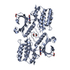 4c58S S: Starting model for refinement |
|---|---|
| Similar structure data |
-Links
 PDBj
PDBj


- Assembly
| Deposited unit | 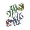
| ||||||||||||||||||||||||||||||||||||||||||||||||||||||||||||||||||||
|---|---|---|---|---|---|---|---|---|---|---|---|---|---|---|---|---|---|---|---|---|---|---|---|---|---|---|---|---|---|---|---|---|---|---|---|---|---|---|---|---|---|---|---|---|---|---|---|---|---|---|---|---|---|---|---|---|---|---|---|---|---|---|---|---|---|---|---|---|---|
| 1 | 
| ||||||||||||||||||||||||||||||||||||||||||||||||||||||||||||||||||||
| 2 | 
| ||||||||||||||||||||||||||||||||||||||||||||||||||||||||||||||||||||
| Unit cell |
| ||||||||||||||||||||||||||||||||||||||||||||||||||||||||||||||||||||
| Noncrystallographic symmetry (NCS) | NCS domain:
NCS domain segments: Component-ID: _ / Refine code: _
NCS ensembles :
|
-Components
| #1: Protein | Mass: 38183.551 Da / Num. of mol.: 2 / Fragment: UNP residues 14-351 Source method: isolated from a genetically manipulated source Source: (gene. exp.) Homo sapiens (human) / Gene: GAK / Plasmid: pNIC28-Bsa4 / Production host:  References: UniProt: O14976, non-specific serine/threonine protein kinase #2: Protein | Mass: 15085.511 Da / Num. of mol.: 2 / Source method: isolated from a natural source / Source: (natural) #3: Chemical |   Mass: 342.415 Da / Num. of mol.: 2 / Source method: obtained synthetically / Formula: C17H18N4O2S Mass: 342.415 Da / Num. of mol.: 2 / Source method: obtained synthetically / Formula: C17H18N4O2S#4: Chemical | ChemComp-EDO /   Mass: 62.068 Da / Num. of mol.: 5 / Source method: obtained synthetically / Formula: C2H6O2 Mass: 62.068 Da / Num. of mol.: 5 / Source method: obtained synthetically / Formula: C2H6O2#5: Water | ChemComp-HOH / |  Mass: 18.015 Da / Num. of mol.: 394 / Source method: isolated from a natural source / Formula: H2O Mass: 18.015 Da / Num. of mol.: 394 / Source method: isolated from a natural source / Formula: H2OHas protein modification | Y | |
|---|
-Experimental details
-Experiment
| Experiment | Method: X-RAY DIFFRACTION |
|---|
- Sample preparation
| Crystal | Density Matthews: 2.01 Å3/Da / Density % sol: 38.75 % |
|---|---|
| Crystal grow | Temperature: 293.15 K / Method: vapor diffusion, sitting drop / pH: 6.2 Details: 9% Broad-molecular weight PEG Smears (BMW PEG smears), 0.1M MES pH 6.2, 0.15M calcium chloride |
-Data collection
| Diffraction | Mean temperature: 100 K |
|---|---|
| Diffraction source | Source: SYNCHROTRON / Site: Diamond  / Beamline: I04-1 / Wavelength: 0.92 Å / Beamline: I04-1 / Wavelength: 0.92 Å |
| Detector | Type: DECTRIS PILATUS 2M / Detector: PIXEL / Date: Oct 31, 2013 |
| Radiation | Monochromator: Si (111) double crystal monochromator / Protocol: SINGLE WAVELENGTH / Monochromatic (M) / Laue (L): M / Scattering type: x-ray |
| Radiation wavelength | Wavelength: 0.92 Å / Relative weight: 1 |
| Reflection | Resolution: 2.1→43.58 Å / Num. obs: 45797 / % possible obs: 94.7 % / Redundancy: 3.7 % / Rmerge(I) obs: 0.071 / Net I/σ(I): 11.9 |
| Reflection shell | Resolution: 2.1→2.21 Å / Redundancy: 3.6 % / Rmerge(I) obs: 0.602 / Mean I/σ(I) obs: 2 / % possible all: 92.4 |
- Processing
| Software |
| ||||||||||||||||||||||||||||||||||||||||||||||||||||||||||||||||||||||||||||||||||||||||||||||||||||||||||||||||||||||||||||||||||||||||||||||||||||||||||||||||||||||||||||||||||||||
|---|---|---|---|---|---|---|---|---|---|---|---|---|---|---|---|---|---|---|---|---|---|---|---|---|---|---|---|---|---|---|---|---|---|---|---|---|---|---|---|---|---|---|---|---|---|---|---|---|---|---|---|---|---|---|---|---|---|---|---|---|---|---|---|---|---|---|---|---|---|---|---|---|---|---|---|---|---|---|---|---|---|---|---|---|---|---|---|---|---|---|---|---|---|---|---|---|---|---|---|---|---|---|---|---|---|---|---|---|---|---|---|---|---|---|---|---|---|---|---|---|---|---|---|---|---|---|---|---|---|---|---|---|---|---|---|---|---|---|---|---|---|---|---|---|---|---|---|---|---|---|---|---|---|---|---|---|---|---|---|---|---|---|---|---|---|---|---|---|---|---|---|---|---|---|---|---|---|---|---|---|---|---|---|
| Refinement | Method to determine structure: MOLECULAR REPLACEMENT Starting model: 4C58 Resolution: 2.1→43.58 Å / Cor.coef. Fo:Fc: 0.953 / Cor.coef. Fo:Fc free: 0.922 / SU B: 12.053 / SU ML: 0.157 / Cross valid method: THROUGHOUT / ESU R: 0.252 / ESU R Free: 0.194 / Stereochemistry target values: MAXIMUM LIKELIHOOD / Details: HYDROGENS HAVE BEEN ADDED IN THE RIDING POSITIONS
| ||||||||||||||||||||||||||||||||||||||||||||||||||||||||||||||||||||||||||||||||||||||||||||||||||||||||||||||||||||||||||||||||||||||||||||||||||||||||||||||||||||||||||||||||||||||
| Solvent computation | Ion probe radii: 0.8 Å / Shrinkage radii: 0.8 Å / VDW probe radii: 1.2 Å / Solvent model: MASK | ||||||||||||||||||||||||||||||||||||||||||||||||||||||||||||||||||||||||||||||||||||||||||||||||||||||||||||||||||||||||||||||||||||||||||||||||||||||||||||||||||||||||||||||||||||||
| Displacement parameters | Biso mean: 70.498 Å2
| ||||||||||||||||||||||||||||||||||||||||||||||||||||||||||||||||||||||||||||||||||||||||||||||||||||||||||||||||||||||||||||||||||||||||||||||||||||||||||||||||||||||||||||||||||||||
| Refine analyze | Luzzati coordinate error obs: 0.342 Å | ||||||||||||||||||||||||||||||||||||||||||||||||||||||||||||||||||||||||||||||||||||||||||||||||||||||||||||||||||||||||||||||||||||||||||||||||||||||||||||||||||||||||||||||||||||||
| Refinement step | Cycle: 1 / Resolution: 2.1→43.58 Å
| ||||||||||||||||||||||||||||||||||||||||||||||||||||||||||||||||||||||||||||||||||||||||||||||||||||||||||||||||||||||||||||||||||||||||||||||||||||||||||||||||||||||||||||||||||||||
| Refine LS restraints |
|