 Movie
Movie Controller

[English] 日本語
 Yorodumi
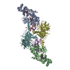
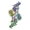
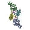
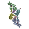
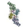
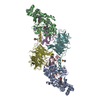
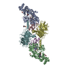
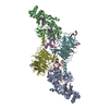
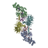
- PDB-4y5r: Crystal Structure of a T67A MauG/pre-Methylamine Dehydrogenase Complex -
+ Open data

- Basic information
| Entry | Database: PDB / ID: 4y5r | ||||||
|---|---|---|---|---|---|---|---|
| Title | Crystal Structure of a T67A MauG/pre-Methylamine Dehydrogenase Complex | ||||||
 Components Components |
| ||||||
 Keywords Keywords | OXIDOREDUCTASE / MauG / Heme / Electron transfer | ||||||
| Function / homology |  Function and homology information Function and homology informationmethylamine dehydrogenase (amicyanin) / methylamine dehydrogenase (amicyanin) activity / methylamine metabolic process / aliphatic amine dehydrogenase activity / amine metabolic process / Oxidoreductases / cytochrome-c peroxidase activity / outer membrane-bounded periplasmic space / periplasmic space / electron transfer activity ...methylamine dehydrogenase (amicyanin) / methylamine dehydrogenase (amicyanin) activity / methylamine metabolic process / aliphatic amine dehydrogenase activity / amine metabolic process / Oxidoreductases / cytochrome-c peroxidase activity / outer membrane-bounded periplasmic space / periplasmic space / electron transfer activity / heme binding / metal ion binding Similarity search - Function | ||||||
| Biological species |  Paracoccus denitrificans (bacteria) Paracoccus denitrificans (bacteria) | ||||||
| Method |  X-RAY DIFFRACTION / SYNCHROTRON / FOURIER SYNTHESIS / Resolution: 2.8 Å X-RAY DIFFRACTION / SYNCHROTRON / FOURIER SYNTHESIS / Resolution: 2.8 Å | ||||||
 Authors Authors | Li, C. / Wilmot, C.M. | ||||||
| Funding support |  United States, 1items United States, 1items
| ||||||
 Citation Citation | Journal: Biochim.Biophys.Acta / Year: 2015 Title: A T67A mutation in the proximal pocket of the high-spin heme of MauG stabilizes formation of a mixed-valent Fe(II)/Fe(III) state and enhances charge resonance stabilization of the bis-Fe(IV) state. Authors: Shin, S. / Feng, M. / Li, C. / Williamson, H.R. / Choi, M. / Wilmot, C.M. / Davidson, V.L. | ||||||
| History |
|
- Structure visualization
| Structure viewer | Molecule: MolmilJmol/JSmol |
|---|
- Downloads & links
-Download
| PDBx/mmCIF format | 4y5r.cif.gz | 656.9 KB | Display | PDBx/mmCIF format |
|---|---|---|---|---|
| PDB format | pdb4y5r.ent.gz | 541.2 KB | Display | PDB format |
| PDBx/mmJSON format | 4y5r.json.gz | Tree view | PDBx/mmJSON format | |
| Others |  Other downloads Other downloads |
-Validation report
| Summary document | 4y5r_validation.pdf.gz | 1.7 MB | Display | wwPDB validaton report |
|---|---|---|---|---|
| Full document | 4y5r_full_validation.pdf.gz | 1.7 MB | Display | |
| Data in XML | 4y5r_validation.xml.gz | 65.6 KB | Display | |
| Data in CIF | 4y5r_validation.cif.gz | 84.8 KB | Display | |
| Arichive directory | https://data.pdbj.org/pub/pdb/validation_reports/y5/4y5rftp://data.pdbj.org/pub/pdb/validation_reports/y5/4y5r | HTTPS FTP |
-Related structure data
| Related structure data | 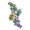 3l4mS S: Starting model for refinement |
|---|---|
| Similar structure data |

-Links
 PDBj
PDBj


- Assembly
| Deposited unit | 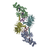
| ||||||||
|---|---|---|---|---|---|---|---|---|---|
| 1 | 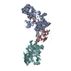
| ||||||||
| Unit cell |
|
-Components
-Protein / Antibody / Methylamine dehydrogenase ... , 3 types, 6 molecules ABCEDF
| #1: Protein | Mass: 38946.277 Da / Num. of mol.: 2 / Fragment: UNP RESIDUES 26-380 / Mutation: T67A Source method: isolated from a genetically manipulated source Source: (gene. exp.) Paracoccus denitrificans (strain Pd 1222) (bacteria)Strain: Pd 1222 / Gene: mauG, Pden_4736 / Production host: Paracoccus denitrificans PD1222 (bacteria) / References: UniProt: Q51658, Oxidoreductases#2: Antibody | Mass: 13714.224 Da / Num. of mol.: 2 / Fragment: UNP RESIDUES 64-188 Source method: isolated from a genetically manipulated source Source: (gene. exp.) Paracoccus denitrificans (bacteria) / Gene: mauA / Production host: Rhodobacter sphaeroides (bacteria)References: UniProt: P22619, methylamine dehydrogenase (amicyanin) #3: Protein | Mass: 41408.266 Da / Num. of mol.: 2 / Fragment: UNP RESIDUES 42-417 Source method: isolated from a genetically manipulated source Source: (gene. exp.) Paracoccus denitrificans (strain Pd 1222) (bacteria)Strain: Pd 1222 / Gene: Pden_4730 / Production host: Rhodobacter sphaeroides (bacteria)References: UniProt: A1BB97, methylamine dehydrogenase (amicyanin) |
|---|
-Non-polymers , 3 types, 12 molecules 


| #4: Chemical |  Mass: 40.078 Da / Num. of mol.: 2 / Source method: obtained synthetically / Formula: Ca Mass: 40.078 Da / Num. of mol.: 2 / Source method: obtained synthetically / Formula: Ca#5: Chemical | ChemComp-HEC /  Mass: 618.503 Da / Num. of mol.: 4 / Source method: obtained synthetically / Formula: C34H34FeN4O4 Mass: 618.503 Da / Num. of mol.: 4 / Source method: obtained synthetically / Formula: C34H34FeN4O4#6: Water | ChemComp-HOH / | Mass: 18.015 Da / Num. of mol.: 6 / Source method: isolated from a natural source / Formula: H2O |
|---|
-Details
| Has protein modification | Y |
|---|
-Experimental details
-Experiment
| Experiment | Method: X-RAY DIFFRACTION / Number of used crystals: 1 |
|---|
- Sample preparation
| Crystal | Density Matthews: 2.38 Å3/Da / Density % sol: 48.37 % |
|---|---|
| Crystal grow | Temperature: 292 K / Method: vapor diffusion, hanging drop / pH: 6.4 Details: 25-30% PEG 8000, 0,1 M sodium Acetate, 0.1 M MES pH 6.4 |
-Data collection
| Diffraction | Mean temperature: 100 K | |||||||||||||||||||||||||||
|---|---|---|---|---|---|---|---|---|---|---|---|---|---|---|---|---|---|---|---|---|---|---|---|---|---|---|---|---|
| Diffraction source | Source: SYNCHROTRON / Site: APS / Beamline: 23-ID-B / Wavelength: 1.003 Å | |||||||||||||||||||||||||||
| Detector | Type: MAR scanner 300 mm plate / Detector: IMAGE PLATE / Date: Oct 23, 2014 | |||||||||||||||||||||||||||
| Radiation | Monochromator: Si / Protocol: SINGLE WAVELENGTH / Monochromatic (M) / Laue (L): M / Scattering type: x-ray | |||||||||||||||||||||||||||
| Radiation wavelength | Wavelength: 1.003 Å / Relative weight: 1 | |||||||||||||||||||||||||||
| Reflection | Resolution: 2.58→29.61 Å / Num. obs: 51683 / % possible obs: 95.2 % / Redundancy: 1.9 % / CC1/2: 0.902 / Rmerge(I) obs: 0.111 / Rpim(I) all: 0.111 / Net I/σ(I): 5.2 / Num. measured all: 98206 | |||||||||||||||||||||||||||
| Reflection shell | Diffraction-ID: 1 / Rejects: _
|
- Processing
| Software |
| |||||||||||||||||||||||||||||||||||||||||||||||||||||||||||||||||||||||||||||||||||||||||||||||||||||||||||||||||||||||||||||||||||||||||||||||||||||||||||||||||||||||||||||||
|---|---|---|---|---|---|---|---|---|---|---|---|---|---|---|---|---|---|---|---|---|---|---|---|---|---|---|---|---|---|---|---|---|---|---|---|---|---|---|---|---|---|---|---|---|---|---|---|---|---|---|---|---|---|---|---|---|---|---|---|---|---|---|---|---|---|---|---|---|---|---|---|---|---|---|---|---|---|---|---|---|---|---|---|---|---|---|---|---|---|---|---|---|---|---|---|---|---|---|---|---|---|---|---|---|---|---|---|---|---|---|---|---|---|---|---|---|---|---|---|---|---|---|---|---|---|---|---|---|---|---|---|---|---|---|---|---|---|---|---|---|---|---|---|---|---|---|---|---|---|---|---|---|---|---|---|---|---|---|---|---|---|---|---|---|---|---|---|---|---|---|---|---|---|---|---|---|
| Refinement | Method to determine structure: FOURIER SYNTHESIS Starting model: 3L4M Resolution: 2.8→29.61 Å / Cor.coef. Fo:Fc: 0.92 / Cor.coef. Fo:Fc free: 0.855 / SU B: 37.461 / SU ML: 0.332 / Cross valid method: THROUGHOUT / σ(F): 0 / ESU R Free: 0.427 / Stereochemistry target values: MAXIMUM LIKELIHOOD Details: HYDROGENS HAVE BEEN ADDED IN THE RIDING POSITIONS U VALUES : WITH TLS ADDED
| |||||||||||||||||||||||||||||||||||||||||||||||||||||||||||||||||||||||||||||||||||||||||||||||||||||||||||||||||||||||||||||||||||||||||||||||||||||||||||||||||||||||||||||||
| Solvent computation | Ion probe radii: 0.8 Å / Shrinkage radii: 0.8 Å / VDW probe radii: 1.2 Å / Solvent model: MASK | |||||||||||||||||||||||||||||||||||||||||||||||||||||||||||||||||||||||||||||||||||||||||||||||||||||||||||||||||||||||||||||||||||||||||||||||||||||||||||||||||||||||||||||||
| Displacement parameters | Biso max: 132.79 Å2 / Biso mean: 51.054 Å2 / Biso min: 26.75 Å2
| |||||||||||||||||||||||||||||||||||||||||||||||||||||||||||||||||||||||||||||||||||||||||||||||||||||||||||||||||||||||||||||||||||||||||||||||||||||||||||||||||||||||||||||||
| Refinement step | Cycle: final / Resolution: 2.8→29.61 Å
| |||||||||||||||||||||||||||||||||||||||||||||||||||||||||||||||||||||||||||||||||||||||||||||||||||||||||||||||||||||||||||||||||||||||||||||||||||||||||||||||||||||||||||||||
| Refine LS restraints |
| |||||||||||||||||||||||||||||||||||||||||||||||||||||||||||||||||||||||||||||||||||||||||||||||||||||||||||||||||||||||||||||||||||||||||||||||||||||||||||||||||||||||||||||||
| LS refinement shell | Resolution: 2.8→2.872 Å / Total num. of bins used: 20
| |||||||||||||||||||||||||||||||||||||||||||||||||||||||||||||||||||||||||||||||||||||||||||||||||||||||||||||||||||||||||||||||||||||||||||||||||||||||||||||||||||||||||||||||
| Refinement TLS params. | Method: refined / Refine-ID: X-RAY DIFFRACTION
| |||||||||||||||||||||||||||||||||||||||||||||||||||||||||||||||||||||||||||||||||||||||||||||||||||||||||||||||||||||||||||||||||||||||||||||||||||||||||||||||||||||||||||||||
| Refinement TLS group |
|
