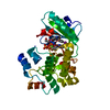
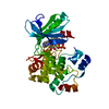
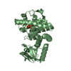
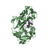
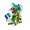
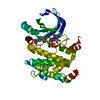
[English] 日本語
Yorodumi
- PDB-4xx9: Crystal structure of PDK1 in complex with ATP and the PIF-pocket ... -

+
Open data


ID or keywords:

Loading...

-
Basic information

Entry
Database: PDB / ID: 4xx9
TitleCrystal structure of PDK1 in complex with ATP and the PIF-pocket ligand RF4
Components3-phosphoinositide-dependent protein kinase 1
KeywordsTransferase/transferase inhibitor / Protein Kinase / Allostery / Transferase-transferase inhibitor complex
Function / homology
Function and homology information


3-phosphoinositide-dependent protein kinase activity / Activation of AKT2 / regulation of mast cell degranulation / negative regulation of toll-like receptor signaling pathway / type B pancreatic cell development / positive regulation of phospholipase activity / RSK activation / hyperosmotic response / regulation of canonical NF-kappaB signal transduction / positive regulation of vascular endothelial cell proliferation ...3-phosphoinositide-dependent protein kinase activity / Activation of AKT2 / regulation of mast cell degranulation / negative regulation of toll-like receptor signaling pathway / type B pancreatic cell development / positive regulation of phospholipase activity / RSK activation / hyperosmotic response / regulation of canonical NF-kappaB signal transduction / positive regulation of vascular endothelial cell proliferation / negative regulation of cardiac muscle cell apoptotic process / phospholipase activator activity / positive regulation of sprouting angiogenesis / Constitutive Signaling by AKT1 E17K in Cancer / CD28 dependent PI3K/Akt signaling / phospholipase binding / positive regulation of blood vessel endothelial cell migration / Role of LAT2/NTAL/LAB on calcium mobilization / Estrogen-stimulated signaling through PRKCZ / SARS-CoV-2 targets host intracellular signalling and regulatory pathways / negative regulation of endothelial cell apoptotic process / SARS-CoV-1 targets host intracellular signalling and regulatory pathways / extrinsic apoptotic signaling pathway / RHO GTPases activate PKNs / cellular response to epidermal growth factor stimulus / GPVI-mediated activation cascade / T cell costimulation / activation of protein kinase B activity / Integrin signaling / insulin-like growth factor receptor signaling pathway / positive regulation of release of sequestered calcium ion into cytosol / VEGFR2 mediated vascular permeability / VEGFR2 mediated cell proliferation / cell projection / calcium-mediated signaling / positive regulation of protein localization to plasma membrane / peptidyl-threonine phosphorylation / negative regulation of transforming growth factor beta receptor signaling pathway / negative regulation of protein kinase activity / epidermal growth factor receptor signaling pathway / CLEC7A (Dectin-1) signaling / FCERI mediated NF-kB activation / G beta:gamma signalling through PI3Kgamma / cellular response to insulin stimulus / positive regulation of angiogenesis / Regulation of TP53 Degradation / cell migration / Downstream TCR signaling / insulin receptor signaling pathway / PIP3 activates AKT signaling / cytoplasmic vesicle / actin cytoskeleton organization / protein autophosphorylation / postsynaptic density / positive regulation of phosphatidylinositol 3-kinase/protein kinase B signal transduction / non-specific serine/threonine protein kinase / intracellular signal transduction / protein phosphorylation / protein serine kinase activity / focal adhesion / protein serine/threonine kinase activity / ATP binding / nucleus / plasma membrane / cytoplasm / cytosol
Similarity search - Function
PDK1-type, PH domain / PDPK1 family / PH domain / PH-like domain superfamily / Transferase(Phosphotransferase) domain 1 / Transferase(Phosphotransferase); domain 1 / Phosphorylase Kinase; domain 1 / Phosphorylase Kinase; domain 1 / Serine/threonine-protein kinase, active site / Serine/Threonine protein kinases active-site signature. ...PDK1-type, PH domain / PDPK1 family / PH domain / PH-like domain superfamily / Transferase(Phosphotransferase) domain 1 / Transferase(Phosphotransferase); domain 1 / Phosphorylase Kinase; domain 1 / Phosphorylase Kinase; domain 1 / Serine/threonine-protein kinase, active site / Serine/Threonine protein kinases active-site signature. / Protein kinase domain / Serine/Threonine protein kinases, catalytic domain / Protein kinase, ATP binding site / Protein kinases ATP-binding region signature. / Protein kinase domain profile. / Protein kinase domain / Protein kinase-like domain superfamily / 2-Layer Sandwich / Orthogonal Bundle / Mainly Alpha / Alpha Beta
Similarity search - Domain/homology
ADENOSINE-5'-TRIPHOSPHATE / Chem-RF4 / 3-phosphoinositide-dependent protein kinase 1
Similarity search - Component
Biological speciesHomo sapiens (human)
MethodX-RAY DIFFRACTION / SYNCHROTRON / MOLECULAR REPLACEMENT / Resolution: 1.4 Å
AuthorsRettenmaier, T.J. / Wells, J.A.
Funding support United States, 1items
OrganizationGrant numberCountry
National Institutes of Health/National Cancer Institute (NIH/NCI)R01 CA136779 United States
CitationJournal: J.Med.Chem. / Year: 2015
Title: Small-Molecule Allosteric Modulators of the Protein Kinase PDK1 from Structure-Based Docking.
Authors: Rettenmaier, T.J. / Fan, H. / Karpiak, J. / Doak, A. / Sali, A. / Shoichet, B.K. / Wells, J.A.
History
DepositionJan 29, 2015Deposition site: RCSB / Processing site: RCSB
Revision 1.0Oct 21, 2015Provider: repository / Type: Initial release
Revision 1.1Nov 4, 2015Group: Database references
Revision 1.2Sep 6, 2017Group: Author supporting evidence / Database references / Derived calculations
Category: citation / pdbx_audit_support / pdbx_struct_oper_list
Item: _citation.journal_id_CSD / _pdbx_audit_support.funding_organization / _pdbx_struct_oper_list.symmetry_operation
Revision 1.3Dec 4, 2019Group: Author supporting evidence / Database references / Category: pdbx_audit_support / struct_ref_seq_dif
Item: _pdbx_audit_support.funding_organization / _struct_ref_seq_dif.details
Revision 1.4Sep 27, 2023Group: Data collection / Database references / Refinement description
Category: chem_comp_atom / chem_comp_bond ...chem_comp_atom / chem_comp_bond / database_2 / pdbx_initial_refinement_model
Item: _database_2.pdbx_DOI / _database_2.pdbx_database_accession

-
Structure visualization

Structure viewerMolecule:
MolmilJmol/JSmol

Downloads & links

-
Assembly

Deposited unit
A: 3-phosphoinositide-dependent protein kinase 1
hetero molecules


Theoretical massNumber of molelcules
Total (without water)36,8327
Polymers35,3361
Non-polymers1,4966
Water4,846269
1


  • Idetical with deposited unit
  • defined by author&software
TypeNameSymmetry operationNumber
identity operation1_555x,y,z1
MethodPISA
Unit cell
Length a, b, c (Å)148.626, 44.429, 47.639
Angle α, β, γ (deg.)90.000, 101.110, 90.000
Int Tables number5
Space group name H-MC121

-
Components

#1: Protein 3-phosphoinositide-dependent protein kinase 1 / hPDK1


Mass: 35335.512 Da / Num. of mol.: 1 / Fragment: UNP residues 50-359 / Mutation: Y288G, Q292A
Source method: isolated from a genetically manipulated source
Source: (gene. exp.) Homo sapiens (human) / Gene: PDPK1, PDK1 / Plasmid: pFastBac HTB / Production host: 
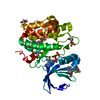
Spodoptera frugiperda (fall armyworm) / Strain (production host): SF9
References: UniProt: O15530, non-specific serine/threonine protein kinase
#2: Chemical ChemComp-ATP / ADENOSINE-5'-TRIPHOSPHATE


Mass: 507.181 Da / Num. of mol.: 1 / Source method: obtained synthetically / Formula: C10H16N5O13P3 / Comment: ATP, energy-carrying molecule*YM
#3: Chemical ChemComp-RF4 / (2-{[2-(2,6-dimethylphenoxy)ethyl]sulfanyl}-1H-benzimidazol-1-yl)acetic acid


Mass: 356.439 Da / Num. of mol.: 2 / Source method: obtained synthetically / Formula: C19H20N2O3S
#4: Chemical ChemComp-GOL / GLYCEROL / GLYCERIN / PROPANE-1,2,3-TRIOL


Mass: 92.094 Da / Num. of mol.: 3 / Source method: obtained synthetically / Formula: C3H8O3
#5: Water ChemComp-HOH / water


Mass: 18.015 Da / Num. of mol.: 269 / Source method: isolated from a natural source / Formula: H2O

-
Experimental details

-
Experiment

ExperimentMethod: X-RAY DIFFRACTION / Number of used crystals: 1

-
Sample preparation

CrystalDensity Matthews: 2.18 Å3/Da / Density % sol: 43.68 %
Crystal growTemperature: 291 K / Method: vapor diffusion, hanging drop / pH: 7.5
Details: Precipitant: 0.1M HEPES, pH 7.5, 1.2M Na Citrate. Protein (21 mg/mL): 25mM Tris, pH 7.5, 0.5M NaCl, 1mM DTT, 15mM EDTA, 15mM ATP

-
Data collection

DiffractionMean temperature: 100 K
Diffraction sourceSource: SYNCHROTRON / Site: ALS / Beamline: 8.3.1 / Wavelength: 1.1159 Å
DetectorType: ADSC QUANTUM 315r / Detector: CCD / Date: Apr 18, 2013
RadiationMonochromator: Double flat crystal, Si 111 / Protocol: SINGLE WAVELENGTH / Monochromatic (M) / Laue (L): M / Scattering type: x-ray
Radiation wavelengthWavelength: 1.1159 Å / Relative weight: 1
ReflectionResolution: 1.4→50 Å / Num. obs: 59563 / % possible obs: 99.7 % / Redundancy: 3.8 % / Biso Wilson estimate: 13.63 Å2 / Rmerge(I) obs: 0.065 / Χ2: 1.112 / Net I/av σ(I): 18.919 / Net I/σ(I): 13.6 / Num. measured all: 225159
Reflection shell

Diffraction-ID: 1 / Rejects: _

Resolution (Å)Redundancy (%)Rmerge(I) obsNum. unique allΧ2% possible all
1.4-1.453.60.82858751.11799.6
1.45-1.513.80.59959451.13699.9
1.51-1.583.80.42159501.15999.9
1.58-1.663.80.30159171.13999.9
1.66-1.763.90.20259511.16399.9
1.76-1.93.90.13559501.10499.8
1.9-2.093.90.08359351.00899.7
2.09-2.393.80.06559581.08299.7
2.39-3.023.70.05559761.10899.6
3.02-503.70.02961061.10199.3

-
Processing

Software
NameVersionClassification
Blu-Icedata collection
PHENIX(phenix.refine: 1.9_1692)refinement
PDB_EXTRACT3.15data extraction
HKL-2000data reduction
HKL-2000data scaling
Coot8model building
RefinementMethod to determine structure: MOLECULAR REPLACEMENT
Starting model: PDB entry 4AW1
Resolution: 1.4→46.746 Å / SU ML: 0.13 / Cross valid method: FREE R-VALUE / σ(F): 1.36 / Phase error: 15.68 / Stereochemistry target values: ML
RfactorNum. reflection% reflection
Rfree0.1651 3010 5.05 %
Rwork0.1286 56551 -
obs0.1304 59561 99.54 %
Solvent computationShrinkage radii: 0.9 Å / VDW probe radii: 1.11 Å / Solvent model: FLAT BULK SOLVENT MODEL
Displacement parametersBiso max: 80.5 Å2 / Biso mean: 22.2821 Å2 / Biso min: 6.12 Å2
Refinement stepCycle: final / Resolution: 1.4→46.746 Å
ProteinNucleic acidLigandSolventTotal
Num. atoms2348 0 124 276 2748
Biso mean--32.31 33.76 -
Num. residues----289
Refine LS restraints
Refine-IDTypeDev idealNumber
X-RAY DIFFRACTIONf_bond_d0.0122573
X-RAY DIFFRACTIONf_angle_d1.4683498
X-RAY DIFFRACTIONf_chiral_restr0.088375
X-RAY DIFFRACTIONf_plane_restr0.008434
X-RAY DIFFRACTIONf_dihedral_angle_d14.975996
LS refinement shell

Refine-ID: X-RAY DIFFRACTION / Total num. of bins used: 22

Resolution (Å)Rfactor RfreeNum. reflection RfreeRfactor RworkNum. reflection RworkNum. reflection all% reflection obs (%)
1.4-1.42560.27251400.2232417255795
1.4256-1.44890.28471230.201525732696100
1.4489-1.47390.2421310.178425462677100
1.4739-1.50070.20571310.158725682699100
1.5007-1.52960.20231300.157925932723100
1.5296-1.56080.21051240.14225612685100
1.5608-1.59470.19131370.132525842721100
1.5947-1.63180.18011530.129925492702100
1.6318-1.67270.19541400.120325512691100
1.6727-1.71790.16651410.112425872728100
1.7179-1.76840.16511410.107225802721100
1.7684-1.82550.14651340.103225352669100
1.8255-1.89080.1491460.100625732719100
1.8908-1.96650.14091370.09825742711100
1.9665-2.0560.13461400.09625672707100
2.056-2.16440.13961400.101525752715100
2.1644-2.30.13591480.105925492697100
2.3-2.47750.15671400.11425962736100
2.4775-2.72680.18081190.1325992718100
2.7268-3.12130.17341360.136125982734100
3.1213-3.93230.17191350.129726172752100
3.9323-46.77230.1511440.15562659280399

+
About Yorodumi

-
News

-
Feb 9, 2022. New format data for meta-information of EMDB entries

New format data for meta-information of EMDB entries

  • Version 3 of the EMDB header file is now the official format.
  • The previous official version 1.9 will be removed from the archive.

Related info.:EMDB header

External links:wwPDB to switch to version 3 of the EMDB data model

-
Aug 12, 2020. Covid-19 info

Covid-19 info

URL: https://pdbjlvh1.pdbj.org/emnavi/covid19.php

New page: Covid-19 featured information page in EM Navigator.

Related info.:Covid-19 info / Mar 5, 2020. Novel coronavirus structure data

+
Mar 5, 2020. Novel coronavirus structure data

Novel coronavirus structure data

Related info.:Yorodumi Speices / Aug 12, 2020. Covid-19 info

External links:COVID-19 featured content - PDBj / Molecule of the Month (242):Coronavirus Proteases

+
Jan 31, 2019. EMDB accession codes are about to change! (news from PDBe EMDB page)

EMDB accession codes are about to change! (news from PDBe EMDB page)

  • The allocation of 4 digits for EMDB accession codes will soon come to an end. Whilst these codes will remain in use, new EMDB accession codes will include an additional digit and will expand incrementally as the available range of codes is exhausted. The current 4-digit format prefixed with “EMD-” (i.e. EMD-XXXX) will advance to a 5-digit format (i.e. EMD-XXXXX), and so on. It is currently estimated that the 4-digit codes will be depleted around Spring 2019, at which point the 5-digit format will come into force.
  • The EM Navigator/Yorodumi systems omit the EMD- prefix.

Related info.:Q: What is EMD? / ID/Accession-code notation in Yorodumi/EM Navigator

External links:EMDB Accession Codes are Changing Soon! / Contact to PDBj

+
Jul 12, 2017. Major update of PDB

Major update of PDB

  • wwPDB released updated PDB data conforming to the new PDBx/mmCIF dictionary.
  • This is a major update changing the version number from 4 to 5, and with Remediation, in which all the entries are updated.
  • In this update, many items about electron microscopy experimental information are reorganized (e.g. em_software).
  • Now, EM Navigator and Yorodumi are based on the updated data.

External links:wwPDB Remediation / Enriched Model Files Conforming to OneDep Data Standards Now Available in the PDB FTP Archive

-
Yorodumi

Thousand views of thousand structures

  • Yorodumi is a browser for structure data from EMDB, PDB, SASBDB, etc.
  • This page is also the successor to EM Navigator detail page, and also detail information page/front-end page for Omokage search.
  • The word "yorodu" (or yorozu) is an old Japanese word meaning "ten thousand". "mi" (miru) is to see.

Related info.:EMDB / PDB / SASBDB / Comparison of 3 databanks / Yorodumi Search / Aug 31, 2016. New EM Navigator & Yorodumi / Yorodumi Papers / Jmol/JSmol / Function and homology information / Changes in new EM Navigator and Yorodumi

Read more