 Movie
Movie Controller


+ Open data

- Basic information
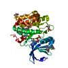
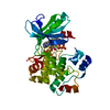
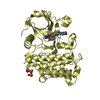
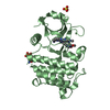
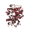
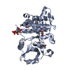
| Entry | Database: PDB / ID: 4rrv | ||||||
|---|---|---|---|---|---|---|---|
| Title | Crystal structure of PDK1 in complex with ATP and PIFtide | ||||||
 Components Components |
| ||||||
 Keywords Keywords | TRANSFERASE/TRANSFERASE INHIBITOR / protein kinase / PIFtide / phosphorylation / TRANSFERASE-TRANSFERASE INHIBITOR complex | ||||||
| Function / homology |  Function and homology information Function and homology informationepithelial cell migration / intracellular signaling cassette / 3-phosphoinositide-dependent protein kinase activity / Activation of AKT2 / regulation of mast cell degranulation / protein kinase C / diacylglycerol-dependent serine/threonine kinase activity / type B pancreatic cell development / negative regulation of toll-like receptor signaling pathway / regulation of cell motility ...epithelial cell migration / intracellular signaling cassette / 3-phosphoinositide-dependent protein kinase activity / Activation of AKT2 / regulation of mast cell degranulation / protein kinase C / diacylglycerol-dependent serine/threonine kinase activity / type B pancreatic cell development / negative regulation of toll-like receptor signaling pathway / regulation of cell motility / apical junction assembly / RSK activation / intermediate filament cytoskeleton / RNA polymerase binding / regulation of canonical NF-kappaB signal transduction / hyperosmotic response / cell projection organization / positive regulation of vascular endothelial cell proliferation / phospholipase activator activity / RHOB GTPase cycle / apical junction complex / positive regulation of sprouting angiogenesis / positive regulation of cytokinesis / RHOC GTPase cycle / Constitutive Signaling by AKT1 E17K in Cancer / cleavage furrow / negative regulation of cardiac muscle cell apoptotic process / Role of LAT2/NTAL/LAB on calcium mobilization / CD28 dependent PI3K/Akt signaling / RHOA GTPase cycle / positive regulation of blood vessel endothelial cell migration / negative regulation of endothelial cell apoptotic process / SARS-CoV-2 targets host intracellular signalling and regulatory pathways / positive regulation of viral genome replication / Estrogen-stimulated signaling through PRKCZ / vascular endothelial cell response to laminar fluid shear stress / SARS-CoV-1 targets host intracellular signalling and regulatory pathways / RHO GTPases activate PKNs / GPVI-mediated activation cascade / phospholipase binding / T cell costimulation / extrinsic apoptotic signaling pathway / Integrin signaling / cell projection / RAC1 GTPase cycle / positive regulation of mitotic cell cycle / insulin-like growth factor receptor signaling pathway / ciliary tip / cellular response to epidermal growth factor stimulus / positive regulation of release of sequestered calcium ion into cytosol / VEGFR2 mediated cell proliferation / VEGFR2 mediated vascular permeability / positive regulation of protein localization to plasma membrane / phosphatidylinositol 3-kinase/protein kinase B signal transduction / negative regulation of transforming growth factor beta receptor signaling pathway / calcium-mediated signaling / CLEC7A (Dectin-1) signaling / small GTPase binding / histone deacetylase binding / epidermal growth factor receptor signaling pathway / FCERI mediated NF-kB activation / cellular response to insulin stimulus / positive regulation of angiogenesis / kinase activity / insulin receptor signaling pathway / Regulation of TP53 Degradation / protein autophosphorylation / PIP3 activates AKT signaling / Downstream TCR signaling / G beta:gamma signalling through PI3Kgamma / cell migration / lamellipodium / actin cytoskeleton organization / protein tyrosine kinase activity / cytoplasmic vesicle / midbody / High laminar flow shear stress activates signaling by PIEZO1 and PECAM1:CDH5:KDR in endothelial cells / protein phosphorylation / protein kinase activity / non-specific serine/threonine protein kinase / positive regulation of phosphatidylinositol 3-kinase/protein kinase B signal transduction / cell adhesion / postsynaptic density / intracellular signal transduction / nuclear body / cadherin binding / protein serine kinase activity / cell division / focal adhesion / protein serine/threonine kinase activity / apoptotic process / centrosome / perinuclear region of cytoplasm / signal transduction / protein-containing complex / RNA binding / nucleoplasm / ATP binding / nucleus / plasma membrane Similarity search - Function | ||||||
| Biological species |  Homo sapiens (human) Homo sapiens (human) | ||||||
| Method |  X-RAY DIFFRACTION / SYNCHROTRON / MOLECULAR REPLACEMENT / Resolution: 1.412 Å X-RAY DIFFRACTION / SYNCHROTRON / MOLECULAR REPLACEMENT / Resolution: 1.412 Å | ||||||
 Authors Authors | Rettenmaier, T.J. / Wells, J.A. | ||||||
 Citation Citation | Journal: Proc.Natl.Acad.Sci.USA / Year: 2014 Title: A small-molecule mimic of a peptide docking motif inhibits the protein kinase PDK1. Authors: Rettenmaier, T.J. / Sadowsky, J.D. / Thomsen, N.D. / Chen, S.C. / Doak, A.K. / Arkin, M.R. / Wells, J.A. | ||||||
| History |
|
- Structure visualization
| Structure viewer | Molecule: MolmilJmol/JSmol |
|---|
- Downloads & links
-Download
| PDBx/mmCIF format | 4rrv.cif.gz | 203.2 KB | Display | PDBx/mmCIF format |
|---|---|---|---|---|
| PDB format | pdb4rrv.ent.gz | 163.2 KB | Display | PDB format |
| PDBx/mmJSON format | 4rrv.json.gz | Tree view | PDBx/mmJSON format | |
| Others |  Other downloads Other downloads |
-Validation report
| Arichive directory | https://data.pdbj.org/pub/pdb/validation_reports/rr/4rrvftp://data.pdbj.org/pub/pdb/validation_reports/rr/4rrv | HTTPS FTP |
|---|
-Related structure data
| Related structure data |  4rqkC  4rqvC  4aw1S C: citing same article ( S: Starting model for refinement |
|---|---|
| Similar structure data |


-Links
 PDBj
PDBj


- Assembly
| Deposited unit | 
| ||||||||
|---|---|---|---|---|---|---|---|---|---|
| 1 |
| ||||||||
| Unit cell |
|
-Components
| #1: Protein | Mass: 35392.566 Da / Num. of mol.: 1 / Fragment: catalytic domain (UNP residues 50-359) / Mutation: Y288G Q292A Source method: isolated from a genetically manipulated source Source: (gene. exp.) Homo sapiens (human) / Gene: PDK1, PDPK1 / Plasmid: pFastBac HTB / Production host:   Spodoptera frugiperda (fall armyworm) / Strain (production host): SF9 Spodoptera frugiperda (fall armyworm) / Strain (production host): SF9References: UniProt: O15530, non-specific serine/threonine protein kinase | ||||
|---|---|---|---|---|---|
| #2: Protein/peptide | Mass: 1995.105 Da / Num. of mol.: 1 Fragment: PIFtide (PRK2 hydrophobic motif, UNP residues 969-983) Source method: obtained synthetically / Source: (synth.) Homo sapiens (human) / References: UniProt: Q16513 | ||||
| #3: Chemical | ChemComp-ATP /   Mass: 507.181 Da / Num. of mol.: 1 / Source method: obtained synthetically / Formula: C10H16N5O13P3 / Comment: ATP, energy-carrying molecule*YM Mass: 507.181 Da / Num. of mol.: 1 / Source method: obtained synthetically / Formula: C10H16N5O13P3 / Comment: ATP, energy-carrying molecule*YM | ||||
| #4: Chemical |   Mass: 92.094 Da / Num. of mol.: 2 / Source method: obtained synthetically / Formula: C3H8O3 Mass: 92.094 Da / Num. of mol.: 2 / Source method: obtained synthetically / Formula: C3H8O3#5: Water | ChemComp-HOH / |  Mass: 18.015 Da / Num. of mol.: 297 / Source method: isolated from a natural source / Formula: H2O Mass: 18.015 Da / Num. of mol.: 297 / Source method: isolated from a natural source / Formula: H2OHas protein modification | Y | |
-Experimental details
-Experiment
| Experiment | Method: X-RAY DIFFRACTION / Number of used crystals: 1 |
|---|
- Sample preparation
| Crystal | Density Matthews: 2.08 Å3/Da / Density % sol: 40.83 % |
|---|---|
| Crystal grow | Temperature: 291 K / Method: vapor diffusion, hanging drop / pH: 7.5 Details: 21 mg/mL protein in 25 mM Tris, pH 7.5, 0.5 M sodium chloride, 1 mM DTT, 15 mM EDTA, 15 mM ATP, precipitant: 0.1 M HEPES, pH 7.5, 1.2 M sodium citrate, VAPOR DIFFUSION, HANGING DROP, temperature 291.0K |
-Data collection
| Diffraction | Mean temperature: 100 K | |||||||||||||||||||||||||||||||||||||||||||||||||||||||||||||||||||||||||||||
|---|---|---|---|---|---|---|---|---|---|---|---|---|---|---|---|---|---|---|---|---|---|---|---|---|---|---|---|---|---|---|---|---|---|---|---|---|---|---|---|---|---|---|---|---|---|---|---|---|---|---|---|---|---|---|---|---|---|---|---|---|---|---|---|---|---|---|---|---|---|---|---|---|---|---|---|---|---|---|
| Diffraction source | Source: SYNCHROTRON / Site: ALS  / Beamline: 8.3.1 / Wavelength: 1.1159 Å / Beamline: 8.3.1 / Wavelength: 1.1159 Å | |||||||||||||||||||||||||||||||||||||||||||||||||||||||||||||||||||||||||||||
| Detector | Type: ADSC QUANTUM 315r / Detector: CCD / Date: Apr 18, 2013 | |||||||||||||||||||||||||||||||||||||||||||||||||||||||||||||||||||||||||||||
| Radiation | Monochromator: Double flat crystal Si(111) / Protocol: SINGLE WAVELENGTH / Monochromatic (M) / Laue (L): M / Scattering type: x-ray | |||||||||||||||||||||||||||||||||||||||||||||||||||||||||||||||||||||||||||||
| Radiation wavelength | Wavelength: 1.1159 Å / Relative weight: 1 | |||||||||||||||||||||||||||||||||||||||||||||||||||||||||||||||||||||||||||||
| Reflection | Resolution: 1.41→50 Å / Num. all: 59080 / Num. obs: 58388 / % possible obs: 98.8 % / Observed criterion σ(F): 0 / Observed criterion σ(I): 0 / Redundancy: 3.5 % / Biso Wilson estimate: 13.81 Å2 / Rmerge(I) obs: 0.061 / Χ2: 1.141 / Net I/σ(I): 13.1 | |||||||||||||||||||||||||||||||||||||||||||||||||||||||||||||||||||||||||||||
| Reflection shell |
|
- Processing
| Software |
| ||||||||||||||||||||||||||||||||||||||||||||||||||||||||||||||||||||||||||||||||||||||||||||||||||||||||||||||||||||||||||||||||||||||||||||||||||||||||||
|---|---|---|---|---|---|---|---|---|---|---|---|---|---|---|---|---|---|---|---|---|---|---|---|---|---|---|---|---|---|---|---|---|---|---|---|---|---|---|---|---|---|---|---|---|---|---|---|---|---|---|---|---|---|---|---|---|---|---|---|---|---|---|---|---|---|---|---|---|---|---|---|---|---|---|---|---|---|---|---|---|---|---|---|---|---|---|---|---|---|---|---|---|---|---|---|---|---|---|---|---|---|---|---|---|---|---|---|---|---|---|---|---|---|---|---|---|---|---|---|---|---|---|---|---|---|---|---|---|---|---|---|---|---|---|---|---|---|---|---|---|---|---|---|---|---|---|---|---|---|---|---|---|---|---|---|
| Refinement | Method to determine structure: MOLECULAR REPLACEMENT Starting model: PDB ENTRY 4AW1 Resolution: 1.412→46.855 Å / SU ML: 0.14 / σ(F): 1.36 / Phase error: 16.19 / Stereochemistry target values: ML
| ||||||||||||||||||||||||||||||||||||||||||||||||||||||||||||||||||||||||||||||||||||||||||||||||||||||||||||||||||||||||||||||||||||||||||||||||||||||||||
| Solvent computation | Shrinkage radii: 0.9 Å / VDW probe radii: 1.11 Å / Solvent model: FLAT BULK SOLVENT MODEL | ||||||||||||||||||||||||||||||||||||||||||||||||||||||||||||||||||||||||||||||||||||||||||||||||||||||||||||||||||||||||||||||||||||||||||||||||||||||||||
| Displacement parameters | Biso max: 88.35 Å2 / Biso mean: 21.8376 Å2 / Biso min: 6.03 Å2 | ||||||||||||||||||||||||||||||||||||||||||||||||||||||||||||||||||||||||||||||||||||||||||||||||||||||||||||||||||||||||||||||||||||||||||||||||||||||||||
| Refinement step | Cycle: LAST / Resolution: 1.412→46.855 Å
| ||||||||||||||||||||||||||||||||||||||||||||||||||||||||||||||||||||||||||||||||||||||||||||||||||||||||||||||||||||||||||||||||||||||||||||||||||||||||||
| Refine LS restraints |
| ||||||||||||||||||||||||||||||||||||||||||||||||||||||||||||||||||||||||||||||||||||||||||||||||||||||||||||||||||||||||||||||||||||||||||||||||||||||||||
| LS refinement shell | Refine-ID: X-RAY DIFFRACTION / Total num. of bins used: 21
|
