 Movie
Movie Controller

[English] 日本語
 Yorodumi
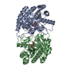
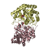
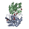
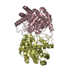
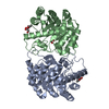
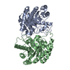
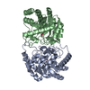
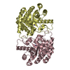
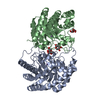
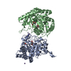
- PDB-4xsm: Crystal structure of D-tagatose 3-epimerase C66S from Pseudomonas... -
+ Open data

- Basic information
| Entry | Database: PDB / ID: 4xsm | ||||||
|---|---|---|---|---|---|---|---|
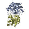
| Title | Crystal structure of D-tagatose 3-epimerase C66S from Pseudomonas cichorii in complex with D-talitol | ||||||
 Components Components | D-tagatose 3-epimerase | ||||||
 Keywords Keywords | ISOMERASE / Epimerase | ||||||
| Function / homology |  Function and homology information Function and homology information | ||||||
| Biological species |  Pseudomonas cichorii (bacteria) Pseudomonas cichorii (bacteria) | ||||||
| Method |  X-RAY DIFFRACTION / MOLECULAR REPLACEMENT / Resolution: 2.3 Å X-RAY DIFFRACTION / MOLECULAR REPLACEMENT / Resolution: 2.3 Å | ||||||
 Authors Authors | Yoshida, H. / Yoshihara, A. / Ishii, T. / Izumori, K. / Kamitori, S. | ||||||
| Funding support |  Japan, 1items Japan, 1items
| ||||||
 Citation Citation | Journal: Appl. Microbiol. Biotechnol. / Year: 2016 Title: X-ray structures of the Pseudomonas cichorii D-tagatose 3-epimerase mutant form C66S recognizing deoxy sugars as substrates Authors: Yoshida, H. / Yoshihara, A. / Ishii, T. / Izumori, K. / Kamitori, S. | ||||||
| History |
|
- Structure visualization
| Structure viewer | Molecule: MolmilJmol/JSmol |
|---|
- Downloads & links
-Download
| PDBx/mmCIF format | 4xsm.cif.gz | 243.2 KB | Display | PDBx/mmCIF format |
|---|---|---|---|---|
| PDB format | pdb4xsm.ent.gz | 194.1 KB | Display | PDB format |
| PDBx/mmJSON format | 4xsm.json.gz | Tree view | PDBx/mmJSON format | |
| Others |  Other downloads Other downloads |
-Validation report
| Summary document | 4xsm_validation.pdf.gz | 490.7 KB | Display | wwPDB validaton report |
|---|---|---|---|---|
| Full document | 4xsm_full_validation.pdf.gz | 527 KB | Display | |
| Data in XML | 4xsm_validation.xml.gz | 50.1 KB | Display | |
| Data in CIF | 4xsm_validation.cif.gz | 68.9 KB | Display | |
| Arichive directory | https://data.pdbj.org/pub/pdb/validation_reports/xs/4xsmftp://data.pdbj.org/pub/pdb/validation_reports/xs/4xsm | HTTPS FTP |
-Related structure data
| Related structure data | 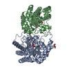 4xslC 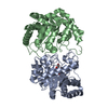 4ytqC 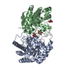 4ytrC  4ytsC  4yttC 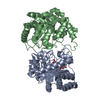 4ytuC 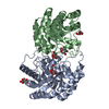 5j8lC 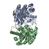 2qulS C: citing same article ( S: Starting model for refinement |
|---|---|
| Similar structure data |


-Links
 PDBj
PDBj
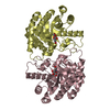
- Assembly
| Deposited unit | 
| ||||||||
|---|---|---|---|---|---|---|---|---|---|
| 1 | 
| ||||||||
| 2 | 
| ||||||||
| Unit cell |
|
-Components
| #1: Protein | Mass: 33858.613 Da / Num. of mol.: 4 / Mutation: C66S Source method: isolated from a genetically manipulated source Source: (gene. exp.) Pseudomonas cichorii (bacteria) / Plasmid: pQE60 / Production host: References: UniProt: O50580, Isomerases; Intramolecular oxidoreductases; Interconverting aldoses and ketoses, and related compounds #2: Chemical | ChemComp-MN /   Mass: 54.938 Da / Num. of mol.: 4 / Source method: obtained synthetically / Formula: Mn Mass: 54.938 Da / Num. of mol.: 4 / Source method: obtained synthetically / Formula: Mn#3: Chemical | ChemComp-TLZ / 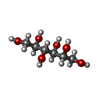  Mass: 182.172 Da / Num. of mol.: 4 / Source method: obtained synthetically / Formula: C6H14O6 Mass: 182.172 Da / Num. of mol.: 4 / Source method: obtained synthetically / Formula: C6H14O6#4: Water | ChemComp-HOH / |  Mass: 18.015 Da / Num. of mol.: 343 / Source method: isolated from a natural source / Formula: H2O Mass: 18.015 Da / Num. of mol.: 343 / Source method: isolated from a natural source / Formula: H2O |
|---|
-Experimental details
-Experiment
| Experiment | Method: X-RAY DIFFRACTION / Number of used crystals: 1 |
|---|
- Sample preparation
| Crystal | Density Matthews: 2.41 Å3/Da / Density % sol: 49.01 % |
|---|---|
| Crystal grow | Temperature: 293 K / Method: vapor diffusion, hanging drop / pH: 4.6 / Details: 6-9% PEG4000, 0.1M Sodium acetate |
-Data collection
| Diffraction | Mean temperature: 100 K | |||||||||||||||||||||||||||||||||||||||||||||||||||||||||||||||||||||||||||||||||||||||||||||||||||
|---|---|---|---|---|---|---|---|---|---|---|---|---|---|---|---|---|---|---|---|---|---|---|---|---|---|---|---|---|---|---|---|---|---|---|---|---|---|---|---|---|---|---|---|---|---|---|---|---|---|---|---|---|---|---|---|---|---|---|---|---|---|---|---|---|---|---|---|---|---|---|---|---|---|---|---|---|---|---|---|---|---|---|---|---|---|---|---|---|---|---|---|---|---|---|---|---|---|---|---|---|
| Diffraction source | Source: ROTATING ANODE / Type: RIGAKU MICROMAX-007 HF / Wavelength: 1.5418 Å | |||||||||||||||||||||||||||||||||||||||||||||||||||||||||||||||||||||||||||||||||||||||||||||||||||
| Detector | Type: RIGAKU RAXIS VII / Detector: IMAGE PLATE / Date: Dec 5, 2014 | |||||||||||||||||||||||||||||||||||||||||||||||||||||||||||||||||||||||||||||||||||||||||||||||||||
| Radiation | Protocol: SINGLE WAVELENGTH / Monochromatic (M) / Laue (L): M / Scattering type: x-ray | |||||||||||||||||||||||||||||||||||||||||||||||||||||||||||||||||||||||||||||||||||||||||||||||||||
| Radiation wavelength | Wavelength: 1.5418 Å / Relative weight: 1 | |||||||||||||||||||||||||||||||||||||||||||||||||||||||||||||||||||||||||||||||||||||||||||||||||||
| Reflection | Resolution: 2.3→25.68 Å / Num. obs: 54404 / % possible obs: 96.7 % / Redundancy: 1.94 % / Biso Wilson estimate: 16.7 Å2 / Rmerge(I) obs: 0.19 / Χ2: 1 / Net I/σ(I): 2.7 / Num. measured all: 106443 / Scaling rejects: 799 | |||||||||||||||||||||||||||||||||||||||||||||||||||||||||||||||||||||||||||||||||||||||||||||||||||
| Reflection shell | Diffraction-ID: 1
|
- Processing
| Software |
| ||||||||||||||||||||||||||||||||||||
|---|---|---|---|---|---|---|---|---|---|---|---|---|---|---|---|---|---|---|---|---|---|---|---|---|---|---|---|---|---|---|---|---|---|---|---|---|---|
| Refinement | Method to determine structure: MOLECULAR REPLACEMENT Starting model: 2QUL Resolution: 2.3→25.68 Å / Rfactor Rfree error: 0.004 / Data cutoff high absF: 3245721 / Data cutoff low absF: 0 / Isotropic thermal model: RESTRAINED / Cross valid method: THROUGHOUT / σ(F): 0 / Details: BULK SOLVENT MODEL USED
| ||||||||||||||||||||||||||||||||||||
| Solvent computation | Solvent model: FLAT MODEL / Bsol: 49.6172 Å2 / ksol: 0.4 e/Å3 | ||||||||||||||||||||||||||||||||||||
| Displacement parameters | Biso max: 83.86 Å2 / Biso mean: 36.1 Å2 / Biso min: 10.52 Å2
| ||||||||||||||||||||||||||||||||||||
| Refine analyze |
| ||||||||||||||||||||||||||||||||||||
| Refinement step | Cycle: final / Resolution: 2.3→25.68 Å
| ||||||||||||||||||||||||||||||||||||
| Refine LS restraints |
| ||||||||||||||||||||||||||||||||||||
| LS refinement shell | Resolution: 2.3→2.38 Å / Rfactor Rfree error: 0.017 / Total num. of bins used: 10
| ||||||||||||||||||||||||||||||||||||
| Xplor file |
|
