 Movie
Movie Controller

[English] 日本語
 Yorodumi
- PDB-4xrf: Crystal structure of MepR like protein complexed with pseudoligands -
+ Open data

- Basic information
| Entry | Database: PDB / ID: 4xrf | ||||||
|---|---|---|---|---|---|---|---|
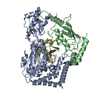
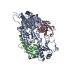
| Title | Crystal structure of MepR like protein complexed with pseudoligands | ||||||
 Components Components | Transcriptional regulator, MarR family | ||||||
 Keywords Keywords | TRANSCRIPTION / WINGED HELIX TURN HELIX TRANSCRIPTION FACTOR | ||||||
| Function / homology |  Function and homology information Function and homology informationresponse to stress / DNA-binding transcription factor activity / regulation of DNA-templated transcription / DNA binding Similarity search - Function | ||||||
| Biological species |  | ||||||
| Method |  X-RAY DIFFRACTION / SYNCHROTRON / MOLECULAR REPLACEMENT / molecular replacement / Resolution: 2.16 Å X-RAY DIFFRACTION / SYNCHROTRON / MOLECULAR REPLACEMENT / molecular replacement / Resolution: 2.16 Å | ||||||
 Authors Authors | Hong, M. / Kim, M.I. / Cho, M.U. | ||||||
 Citation Citation | Journal: to be published Title: Crystal structure of MepR like protein complexed with pseudoligands Authors: Hong, M. / Kim, M.I. / Cho, M.U. | ||||||
| History |
|
- Structure visualization
| Structure viewer | Molecule: MolmilJmol/JSmol |
|---|
- Downloads & links
-Download
| PDBx/mmCIF format | 4xrf.cif.gz | 77.9 KB | Display | PDBx/mmCIF format |
|---|---|---|---|---|
| PDB format | pdb4xrf.ent.gz | 57.7 KB | Display | PDB format |
| PDBx/mmJSON format | 4xrf.json.gz | Tree view | PDBx/mmJSON format | |
| Others |  Other downloads Other downloads |
-Validation report
| Arichive directory | https://data.pdbj.org/pub/pdb/validation_reports/xr/4xrfftp://data.pdbj.org/pub/pdb/validation_reports/xr/4xrf | HTTPS FTP |
|---|
-Related structure data
| Similar structure data |
|---|
-Links
 PDBj
PDBj
- Assembly
| Deposited unit | 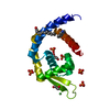
| ||||||||||||
|---|---|---|---|---|---|---|---|---|---|---|---|---|---|
| 1 | 
| ||||||||||||
| Unit cell |
| ||||||||||||
| Components on special symmetry positions |
| ||||||||||||
| Details | The biological assembly is a hexamer generated from the dimer in the asymmetric unit by the operations: -X+Y, Y, -Z |
-Components
-Protein , 1 types, 1 molecules A
| #1: Protein | Mass: 16531.305 Da / Num. of mol.: 1 / Fragment: UNP residues 5-146 Source method: isolated from a genetically manipulated source Source: (gene. exp.) Strain: ATCC 14579 / DSM 31 / Gene: BC_0657 / Plasmid: pET49b / Production host: |
|---|
-Non-polymers , 7 types, 76 molecules 


| #2: Chemical | ChemComp-PO4 /  Mass: 94.971 Da / Num. of mol.: 4 / Source method: obtained synthetically / Formula: PO4 Mass: 94.971 Da / Num. of mol.: 4 / Source method: obtained synthetically / Formula: PO4#3: Chemical | ChemComp-ISQ / |  Mass: 129.159 Da / Num. of mol.: 1 / Source method: obtained synthetically / Formula: C9H7N / Comment: alkaloid*YM Mass: 129.159 Da / Num. of mol.: 1 / Source method: obtained synthetically / Formula: C9H7N / Comment: alkaloid*YM#4: Chemical | ChemComp-DAO / |  Mass: 200.318 Da / Num. of mol.: 1 / Source method: obtained synthetically / Formula: C12H24O2 Mass: 200.318 Da / Num. of mol.: 1 / Source method: obtained synthetically / Formula: C12H24O2#5: Chemical |  Mass: 92.094 Da / Num. of mol.: 2 / Source method: obtained synthetically / Formula: C3H8O3 Mass: 92.094 Da / Num. of mol.: 2 / Source method: obtained synthetically / Formula: C3H8O3#6: Chemical | ChemComp-NI / |  Mass: 58.693 Da / Num. of mol.: 1 / Source method: obtained synthetically / Formula: Ni Mass: 58.693 Da / Num. of mol.: 1 / Source method: obtained synthetically / Formula: Ni#7: Chemical | ChemComp-SO4 / |  Mass: 96.063 Da / Num. of mol.: 1 / Source method: obtained synthetically / Formula: SO4 Mass: 96.063 Da / Num. of mol.: 1 / Source method: obtained synthetically / Formula: SO4#8: Water | ChemComp-HOH / | Mass: 18.015 Da / Num. of mol.: 66 / Source method: isolated from a natural source / Formula: H2O |
|---|
-Experimental details
-Experiment
| Experiment | Method: X-RAY DIFFRACTION / Number of used crystals: 1 |
|---|
- Sample preparation
| Crystal | Density Matthews: 3.63 Å3/Da / Density % sol: 66.16 % |
|---|---|
| Crystal grow | Temperature: 291 K / Method: vapor diffusion, hanging drop / pH: 4 / Details: 0.1M phosphate citrate, 2.4 M ammonium sulfate |
-Data collection
| Diffraction | Mean temperature: 100 K | ||||||||||||||||||||||||||||||||||||||||||||||||||||||||||||||||||||||||||||||||||||||||||||||||||||||||||||||||||||||||||||||
|---|---|---|---|---|---|---|---|---|---|---|---|---|---|---|---|---|---|---|---|---|---|---|---|---|---|---|---|---|---|---|---|---|---|---|---|---|---|---|---|---|---|---|---|---|---|---|---|---|---|---|---|---|---|---|---|---|---|---|---|---|---|---|---|---|---|---|---|---|---|---|---|---|---|---|---|---|---|---|---|---|---|---|---|---|---|---|---|---|---|---|---|---|---|---|---|---|---|---|---|---|---|---|---|---|---|---|---|---|---|---|---|---|---|---|---|---|---|---|---|---|---|---|---|---|---|---|---|
| Diffraction source | Source: SYNCHROTRON / Site: PAL/PLS  / Beamline: 7A (6B, 6C1) / Wavelength: 0.97958 Å / Beamline: 7A (6B, 6C1) / Wavelength: 0.97958 Å | ||||||||||||||||||||||||||||||||||||||||||||||||||||||||||||||||||||||||||||||||||||||||||||||||||||||||||||||||||||||||||||||
| Detector | Type: ADSC QUANTUM 270 / Detector: CCD / Date: Apr 8, 2014 / Details: certical focusing toroidal, rhodium coated | ||||||||||||||||||||||||||||||||||||||||||||||||||||||||||||||||||||||||||||||||||||||||||||||||||||||||||||||||||||||||||||||
| Radiation | Monochromator: DCM Si crystal / Protocol: SINGLE WAVELENGTH / Monochromatic (M) / Laue (L): M / Scattering type: x-ray | ||||||||||||||||||||||||||||||||||||||||||||||||||||||||||||||||||||||||||||||||||||||||||||||||||||||||||||||||||||||||||||||
| Radiation wavelength | Wavelength: 0.97958 Å / Relative weight: 1 | ||||||||||||||||||||||||||||||||||||||||||||||||||||||||||||||||||||||||||||||||||||||||||||||||||||||||||||||||||||||||||||||
| Reflection | Resolution: 2.16→50 Å / Num. obs: 13502 / % possible obs: 99.1 % / Redundancy: 20.6 % / Rmerge(I) obs: 0.094 / Χ2: 5.479 / Net I/av σ(I): 83.969 / Net I/σ(I): 16.8 / Num. measured all: 277710 | ||||||||||||||||||||||||||||||||||||||||||||||||||||||||||||||||||||||||||||||||||||||||||||||||||||||||||||||||||||||||||||||
| Reflection shell | Diffraction-ID: 1 / Rejects: _
|
-Phasing
| Phasing | Method: molecular replacement |
|---|
- Processing
| Software |
| |||||||||||||||||||||||||||||||||||||||||||||||||||||||||||||||||||||||||||
|---|---|---|---|---|---|---|---|---|---|---|---|---|---|---|---|---|---|---|---|---|---|---|---|---|---|---|---|---|---|---|---|---|---|---|---|---|---|---|---|---|---|---|---|---|---|---|---|---|---|---|---|---|---|---|---|---|---|---|---|---|---|---|---|---|---|---|---|---|---|---|---|---|---|---|---|---|
| Refinement | Method to determine structure: MOLECULAR REPLACEMENT / Resolution: 2.16→26.72 Å / Cor.coef. Fo:Fc: 0.944 / Cor.coef. Fo:Fc free: 0.932 / SU B: 9.708 / SU ML: 0.133 / Cross valid method: THROUGHOUT / σ(F): 0 / ESU R: 0.193 / ESU R Free: 0.167 / Stereochemistry target values: MAXIMUM LIKELIHOOD / Details: U VALUES : WITH TLS ADDED
| |||||||||||||||||||||||||||||||||||||||||||||||||||||||||||||||||||||||||||
| Solvent computation | Ion probe radii: 0.8 Å / Shrinkage radii: 0.8 Å / VDW probe radii: 1.2 Å / Solvent model: MASK | |||||||||||||||||||||||||||||||||||||||||||||||||||||||||||||||||||||||||||
| Displacement parameters | Biso max: 97.63 Å2 / Biso mean: 45.695 Å2 / Biso min: 20.67 Å2
| |||||||||||||||||||||||||||||||||||||||||||||||||||||||||||||||||||||||||||
| Refinement step | Cycle: final / Resolution: 2.16→26.72 Å
| |||||||||||||||||||||||||||||||||||||||||||||||||||||||||||||||||||||||||||
| Refine LS restraints |
| |||||||||||||||||||||||||||||||||||||||||||||||||||||||||||||||||||||||||||
| LS refinement shell | Resolution: 2.162→2.219 Å / Total num. of bins used: 20
| |||||||||||||||||||||||||||||||||||||||||||||||||||||||||||||||||||||||||||
| Refinement TLS params. | Method: refined / Refine-ID: X-RAY DIFFRACTION
| |||||||||||||||||||||||||||||||||||||||||||||||||||||||||||||||||||||||||||
| Refinement TLS group |
|
